Aortic Valve Replacement CVS173
Aortic Valve Replacement Transcript
Aortic Valve Replacement
This is Dr. Cal Shipley with a review of aortic valve replacement. In this presentation, I’m going to be depicting replacement of the aortic valve with a bioprosthetic artificial valve.
Bioprosthetic Aortic Valve: Structure
A bioprosthetic valve is so named because the leaflets of the valve are composed of organic animal tissue, most commonly sourced from cows or pigs. The leaflets are attached to a support, also known as a stent, made of metal or a plastic polymer.
Struts
Typically, the support has three struts. The struts allow attachment of the leaflets to the support, so that the movement of the leaflets during opening and closure closely mimics those of a natural biological valve. By closely approximating leaflet movement to those of a biological valve, a bioprosthetic valve allows for a more natural flow of blood through the valve than a mechanical artificial valve. Because blood flow is more natural and less turbulent than it is through a mechanical valve, there is less chance of blood clot formation. This is one of the major advantages of a bioprosthetic tissue valve.
Sewing Ring
Attached around the outer edge of the support is a sewing ring. This is a soft, pliable structure, typically made of polyester, and is used to attach the valve with suture material to the annulus of the aorta.
Relevant Cardiac Anatomy
Now let’s take a look at some of the anatomy involved with aortic valve replacement. The aorta is attached to the top of the left ventricle and receives freshly oxygenated blood for distribution to all bodily organs and tissues.
The aortic annulus is a fibrous ring which marks the border between the left ventricle and the aortic root. It is the point at which the base of each of the three leaflets of the aortic valve attach, and it is considered to be a virtual ring in that it is not continuous and exists only at the point of attachment of the leaflets. Just as it does for the leaflets of the biological aortic valve, the annulus provides a sturdy anchor for the bioprosthetic valve.
Bioprosthetic Valve: Insertion Technique
Let’s take a look now at the insertion technique for the bioprosthetic aortic valve.
There are approximately 180,000 valve replacement surgeries performed in the US each year. This number is split about evenly between mitral valve replacements and aortic valve replacements. While the number of transcatheter valve replacements are increasing each year, the majority of valve replacements are still done by an open technique.
Open Valve Replacement
The open technique requires opening the front of the chest wall in order to access the heart.
Anatomy Encountered with Open Technique Valve Replacement
To better understand the open technique, let’s do a quick review of the relevant anatomy as encountered by the cardiovascular surgeon.
Beneath the skin layers of the chest lies the rib cage with the sternum, also known as the breastbone, at its center. Deep to the rib cage lies the heart, bookended by the lungs and surrounded by its protective membrane, the pericardium. Viewing the heart in cross-section reveals the main pulmonary artery, a portion of which lies directly in front of the aortic root and valve. In order to reach the aortic valve, all of the previously mentioned anatomical structures must be either penetrated or bypassed.
Surgical Procedure
Median Sternotomy
Once the patient has been successfully placed under a general anesthetic, the surgeon begins by making a skin incision over the sternum. Using surgical instruments, the sternum is divided in the midline, a procedure known as a median sternotomy. The sternotomy allows the ribcage and lungs to be retracted to either side, exposing the pericardium. The pericardium is then opened to expose the heart.
Ministernotomy
A recently developed alternative to the traditional sternotomy is the mini-sternotomy. In the mini-sternotomy technique, only the upper half of the sternum is opened with a T-type incision as shown here. As a result, there is less chest wall trauma with the mini-sternotomy. However, operating times tend to be longer due to the restricted access compared to a full sternotomy. So far, studies have yet to show a convincing advantage for patients in the long term.
Cardiopulmonary Bypass
Once the heart has been fully exposed, the patient is placed on cardiopulmonary bypass, also known as a heart-lung machine. The cardiopulmonary bypass system takes the place of the heart and lungs throughout the procedure, oxygenating blood and circulating it through the vascular system. To see more information on how cardiopulmonary bypass works, please take a look at my video located in the cardiovascular system library.
Cardioplegia
Once the patient has been placed on cardiopulmonary bypass, the heart is brought into a state of full cardiac arrest via the administration of a cardioplegia solution, typically potassium-based, into the bloodstream. The cardioplegia solution may be administered throughout the procedure, either by continuous or intermittent infusion or, for shorter procedures, in a single dose.
Removal of the Biological Valve
Once the heart has been arrested, a cross clamp is applied to prevent the backflow of blood from the aorta during the procedure.
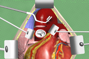
The root of the aorta is exposed as it emerges from the left ventricle. The aorta is then surgically transected, so that the leaflets of the aortic valve may be accessed. The aortic valve leaflets are removed, and the aortic root is now ready to be prepared for the insertion of the bioprosthetic valve. Here is a view looking down into the aortic root after the aortic valve leaflets have been removed and the annulus exposed.
Attachment to the Aortic Annulus
The sutures, which will anchor the valve in place, are placed circumferentially around the annulus. The sutures have a needle on both ends, and these are used to thread the sutures through the sewing ring of the valve. The valve is then repositioned down to the annulus. Each suture, in turn, is tied off to anchor the valve.
Positioning of the Artificial Valve
With this type of artificial valve, it is critically important that placement of the valve is such that none of the struts block the coronary ostiae.
The coronary ostiae are openings in the root of the aorta which allow oxygenated blood to flow into the coronary arteries. Obstruction of an ostia by the strut of an incorrectly placed valve can severely impair coronary artery blood flow, resulting in myocardial ischemia, or even myocardial infarction.
Reinitiating Cardiac Chamber Contraction
Once the bioprosthetic aortic valve has been sewn in place, the previously transected aorta may be closed. Once the aorta has been closed, the cross clamp is removed.
The removal of the aortic cross clamp allows blood to flow into the coronary arteries, and this usually stimulates spontaneous resumption of the heartbeat. Occasionally, a mild electrical shock applied to the heart is required to reinitiate cardiac chamber contraction.
Removal of Cardiopulmonary Bypass and Final Steps
Once the cardiac chambers are contracting normally, the patient is taken off cardiopulmonary bypass. The pericardium, sternum, and skin are closed, and the procedure is complete.
Cal Shipley, M.D. copyright 2020

