Cardiac Ablation for EAT
This is Dr. Cal Shipley with a review of cardiac ablation to treat ectopic atrial tachycardia.
Cardiac Electrophysiology
Let’s start by taking a look at the normal electrophysiology of the heart.
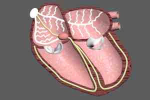
The heart consists of four contracting chambers; the right and left atrium, and the right and left ventricle. The sinoatrial node is a bundle of cells located in the wall of the right atrium. The SA node starts each heartbeat by spontaneously producing an electrical impulse. The impulse travels along the cardiac conduction system, a series of nerve bundles, and spreads throughout the atria, causing them to contract. At the same time, an impulse travels to the atrioventricular node located at the junction between the right atrium and the right ventricle. This, in turn, stimulates an impulse in the atrioventricular node.
The AV node impulse travels along conduction pathways to both ventricles. The impulse spreads through the ventricles and causes them to contract.
This cycle repeats about 60 to 100 times per minute. Normal impulse generation, conduction and heartbeat are known as normal sinus rhythm (NSR).
Ectopic Atrial Tachycardia (EAT)
Let’s take a look at one abnormal manifestation of cardiac electrophysiology; ectopic atrial tachycardia (EAT).
Ectopic Pacemakers
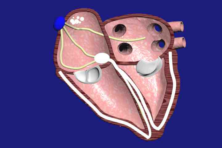
In our review of cardiac electrophysiology, we noted that normal sinus rhythm starts with the sinoatrial node. In ectopic atrial tachycardia, an abnormal cluster of cells located in either the left or right atrium generates spontaneous electrical impulses independently of the SA node. These abnormal impulses are termed ectopic because they are generated in a location which is outside of the normal cardiac conduction system.
The abnormal cluster of cells is designated as an ectopic pacemaker. The ectopic impulses are conducted to the AV node. The ectopic impulses are generated at an abnormally high rate, typically 100 to 250 times per minute.
AV Node Block
A healthy AV node will transmit the impulses from the ectopic pacemaker at a rate no greater than 140 to 150 times per minute. Beyond this point, the AV node will begin to block some of the ectopic pacemaker impulses in order to prevent a dangerously rapid ventricular rate.
Even in the presence of AV node block, ventricular rates of 120 to 150 beats per minute are not uncommon during bursts of ectopic activity. Ectopic atrial tachycardia is one form of supraventricular tachycardia (SVT), that is, tachycardia originating because of electrical abnormalities outside of the ventricles.
EAT: Adverse Effects
In the short term, EAT can precipitate heart failure in individuals with pre-existing cardiac disease. For example, a history of myocardial infarction.
In the long term, the tachycardia associated with recurrent or persistent bouts of EAT can result in cardiomyopathy, with weakening and enlargement of the atria and ventricles.
Cardiac Ablation Procedure for EAT
Now that we have a better understanding of what ectopic atrial tachycardia is, how it occurs, and some of the dangers associated with it, let’s turn to treatment with cardiac ablation.
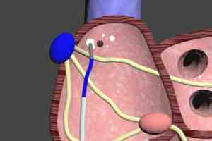
I’m going to use the same example of a cluster of cells generating impulses in the right atrium, but these clusters may occur in either atrium, and in some cases, multiple clusters may be present in either or both atria.
Ablation Catheter Placement
To begin the procedure, a sheath is inserted through either the right or left femoral vein, and then up through the inferior vena cava and into the right atrium. The sheath is a relatively rigid tube which provides a good anchor for the more flexible ablation catheter.
An ablation catheter is then inserted through the sheath. In addition to performing ablation, the catheter is also capable of mapping the ectopic cell clusters so that the electrophysiologist can accurately determine their location prior to, and during, the ablation procedure.
Cardiac Ablation Challenges
Cardiac ablation requires tremendous skill and perseverance on the part of the electrophysiologist. Not the least of the challenges is the fact that the catheter must be accurately manipulated while the heart is beating. In some cases, the procedure may require several hours to complete.
Radio Frequency (RF) Pulses
Once the ectopic cell cluster has been located, radio frequency pulses are emitted, which heat the tissue and ablate the cells.
A typical ectopic cluster will require several passes of the ablation catheter to destroy all of the abnormal cells. This requires several precise positional changes of the catheter tip. As the procedure proceeds, the catheter is used as a mapping tool to determine the effectiveness of the ablations. This process is repeated until all of the ectopic cells have been effectively ablated.
Ablation Success Criteria
The success of the ablation procedure is based on re-establishment of normal sinus rhythm and the elimination of future recurrences of EAT.
Cal Shipley, M.D. copyright 2020