Coronary Artery Perfusion CVS009
Helpful Links
Coronary Artery Perfusion Transcript
Coronary Artery Perfusion
This is Dr. Cal Shipley with a review of left ventricular function and the cardiac cycle in coronary artery perfusion.
The Left Ventricle
The heart is located centrally in the chest cavity, also known as the thorax, and is nestled between the lungs. Among the chambers of the heart, the left ventricle does the lion’s share of the work, pumping freshly oxygenated blood via the aortic valve into the aorta, and from the aorta, blood flows to all bodily tissues and organs.
The Cardiac Cycle
The cardiac cycle consists of the expansion and contraction of the various heart chambers with each heartbeat.
Systole
During the portion of the cardiac cycle known as systole, the left ventricle contracts, pushing blood against aortic valve. This causes the valve to open allowing blood to flow into the aorta.
Diastole
During diastole, the left ventricle relaxes and is refilled with freshly oxygenated blood from the lungs. The aortic valve closes preventing backflow of blood from the aorta.
The term cardiac output refers to the volume of blood pushed from the left ventricle into the aorta with each contraction.
Looking at a view of the heart from the patient’s left side, we can see the aorta and left ventricle in cross section as well as the aortic valve.
From this viewpoint, we can again note the opening of the aortic valve during systole to allow blood flow into the aorta and closure of the valve during diastole to prevent backflow.
The Coronary Arteries
The coronary arteries are responsible for conducting nutrient-rich blood to the heart muscle, also known as the myocardium.
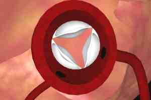
The coronary arteries are attached to the aorta. Changing to a cross-sectional view of the aorta viewed from above, the opening and closing of the aortic valve during the cardiac cycle can be seen, as well as the coronary artery ostia. The coronary ostia are openings in the aortic wall which communicate with the coronary arteries.
Coronary Artery Perfusion
Myocardial Extravascular Compression
As previously noted, during systole, blood is ejected from the left ventricle through the aortic valve and into the aorta. Though it may seem somewhat paradoxical, only a small amount of blood flows into the coronary arteries during systole. The reason for this limited volume of coronary artery blood flow during systole is a phenomenon known as myocardial extravascular compression. Contraction of the myocardium during systole compresses the dense network of arterioles and capillaries that are interwoven throughout the muscle.
This compression of vascular structures causes a marked increase in resistance to blood flow through the coronary system and hence a small volume of flow into the coronary arteries during systole.
During diastole, the myocardium relaxes and the compression of the vascular structures is relieved. The blood pressure in the aortic root is now greater than that of the coronaries and blood flows freely into the ostia through the coronary system.
Summary
To summarize, blood flows into the coronary arteries after flowing across the aortic valve, due to the phenomenon of myocardial extravascular compression. Blood flow through the coronary artery system is highest during diastole and lowest during systole. The heart, then, is responsible for supplying nutrients to itself, as well as to all other bodily tissues and organs.
Cal Shipley, M.D. copyright 2021

