Laparoscopic Appendectomy GST404
Laparoscopic Appendectomy Transcript
Laparoscopic Appendectomy
This is Dr. Cal Shipley with a review of laparoscopic appendectomy.
Abdominal Anatomy
Let’s start with a quick review of the relevant anatomy. The midsection of the abdomen is largely occupied by the small intestine. Running mostly peripherally to the small intestine is the colon. The cecum is a slight outpouching located at the start of the ascending colon on the right side of the abdomen. Attached to the cecum is the appendix.
The mesoappendix is a fold of peritoneum which wraps around the appendix and carries the appendiceal artery and blood supply. The teniae coli are ribbons of smooth muscle which run longitudinally along the outside of the colon. Only one of the teniae can be seen from this viewpoint. The significance of the teniae coli is that they come together and meet at the base of the appendix. As such, they can provide an excellent marker for the orientation of the appendix during appendectomy.
Laparoscopic Appendectomy Procedure
Abdominal Insufflation
The first step in the procedure is to insert a thin needle-like cannula into the abdomen in the area of the umbilicus. Viewing the abdomen from the left side in a cross-sectional view, we can see the cannula inserted into the peritoneal space. In a process known as insufflation, carbon dioxide gas is passed through the cannula into the peritoneal space. Insufflation increases the workspace within the abdominal cavity for the surgeon to manipulate instruments during the procedure.
Abdominal Incisions: Port Creation
Once insufflation of the abdomen has been accomplished, the surgical incisions are made.
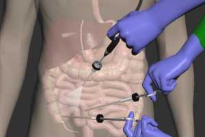
In contrast to open appendectomy, where a 3 to 5-inch incision is made directly over the infected appendix, laparoscopic appendectomy relies on several small quarter to half-inch incisions, all of them made away from the area of appendiceal infection. These small incisions are known as ports.
Trocar Placement
Trocars consisting of cylindrical metal tubes with collars are inserted through the ports. Although there are several variations, the setup of ports and trocars I have depicted here is fairly typical, with placement in the periumbilical region, the left lower quadrant of the abdomen, and the suprapubic area. The left lower quadrant and suprapubic ports are used for surgical instruments. One of the instrument ports may be used for a suction tool to remove blood and excess fluid from the operative field as needed.
The periumbilical port holds the laparoscopic camera which provides the surgeon and her assistants with a view of the operative field throughout the procedure. Operating room video monitors display the images generated by the laparoscopic camera.
Location and Removal of the Appendix
Upon entering the abdominal cavity, the surgeon locates the cecum. Depending on the degree of inflammatory reaction around the infected appendix, a fair amount of tissue dissection may be required to expose the cecum. Once the cecum has been identified, the teniae coli may be used to determine the location of the base of the appendix. Surgical instruments are used to remove fatty and inflammatory tissue around the base of the appendix. Once the base of the appendix has been exposed, the surgeon systematically exposes the rest of the organ to its tip. At the completion of this dissection, the appendix can now be fully visualized.
In this example, about 75% of the appendix has become infected and inflamed. About 25% of the organ, including the base, is normal. Having identified and exposed the appendix, the next step is to separate the appendix from its blood supply. In order to accomplish this, it is necessary to fully expose the mesoappendix which carries the appendiceal artery and its tributaries. Typically, a blunt instrument is used to create a window or hole in the mesoappendix membrane. A Bovie electrocautery unit is then used to cut the membrane, coagulating any vessels it encounters as it moves along. Once the blood supply has been separated, the surgeon confirms the location of the appendiceal base using the junction of the three teniae coli as an anatomical landmark.
A surgical stapler is positioned over the appendecial base, and with one action, it severs the base from the cecum and also staples and seals both sides of the incision. The remaining appendiceal stump is about 5 millimeters in length. The infected appendix is now free of all attachments and may be removed.
Cal Shipley, M.D. copyright 2020

