Peritonitis Transcript
Peritonitis
This is Dr. Cal Shipley with a review of perforated stomach ulcer and acute peritonitis.
Abdominal Anatomy
Let’s do a quick review of the abdominal anatomy including the liver, the stomach, and the small intestine. The jejunum, which is the proximal half of the intestine, lies beneath the titles here, but the entire small intestine, including the distal portion, also known as the ileum, is pictured. And the colon, also known as the large intestine, and the peritoneum, or peritoneal membrane, which enfolds and covers most of the stomach, large and small intestines, and liver. The peritoneal membrane blankets the abdominal organs, acting as a barrier to infection and trauma. It also holds the organs in place, particularly the intestines, and prevents them from twisting or kinking. The peritoneum also carries the complex web of blood vessels to and from the organs. The large fold of the peritoneal membrane hangs like an apron from the margins of the transverse colon, and is known as the greater omentum.
Gastric ulcers erode the inner lining of the stomach. If untreated, such ulcers may eventually create a through and through perforation. Once perforation occurs, there is nothing to stop stomach contents from leaking into the open abdominal cavity. This leakage may consist of food, stomach acid, bacteria, and bile, all of which may have a very toxic, irritating effect on the peritoneal membranes.
Acute Peritonitis
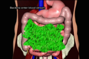
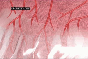
The peritoneum becomes inflamed and may become infected. This is known as acute peritonitis. The underlying organs also become inflamed. Exudates and adhesive bands may form among the organs. Ongoing inflammation may result in compromise of blood flow to the organs, resulting in necrosis. Bacteria may cause infection which may enter the blood stream, causing septicemia and possibly septic shock.
Please see my animation on septic shock in this directory as well as the shock directory for more information.
Cal Shipley, M.D. copyright 2020


