DIEP Breast Reconstruction Transcript
Relevant Anatomy in DIEP Breast Reconstruction
Abdominal Wall Anatomy
This is Doctor Cal Shipley with a review of the DIEP breast reconstruction procedure. DIEP is an acronym for Deep Inferior Epigastric Perforator arteries. Let’s take a look at the anatomy of the abdominal wall. Just beneath the skin layers, even in lean individuals, lies a fairly thick fat pad which covers the entirety of the abdomen. The fascia helps to stabilize the muscle’s position within the abdomen, and also makes it easier for muscles to work in concert.
In this image, I have highlighted these sheets surrounding the rectus abdominis muscles. Here we see the rectus abdominus muscles with the fascia removed.
Vascular Anatomy
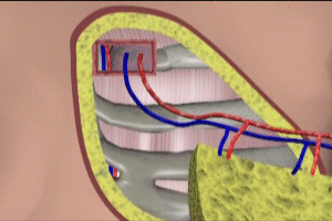
Now, let’s take a look at the blood supply to the rectus muscles. Blood flow to the rectus muscles arises from the deep inferior epigastric arteries, which themselves arise from the iliac arteries in the pelvis. The deep inferior epigastric does not only supply blood to the rectus muscles but also to the overlying fat layer and skin of the abdominal wall.
The other key vascular structures involved in the deep breast reconstruction procedure are the internal thoracic artery and vein. The internal thoracic vessels are located in the thorax or chest cavity, and lie just to the side of the sternum. Depicted here are the left internal thoracic vein and artery. These vessels also lie very close to the anterior chest wall and this location is critical to their usefulness in the deep procedure.
DIEP Breast Reconstruction Surgical Technique
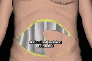
The abdominal skin incision for the deep procedure is elliptical. A full ellipse if doing a bilateral breast reconstruction, and half an ellipse if doing a unilateral reconstruction. Once the incision is made, a skin island consisting of the epidermal, dermal, and subcutaneous fat layers is elevated. The deep inferior epigastric perforator arteries are then inspected. It is critical to the success of the deep procedure that the perforator arteries be of adequate size. Perforator artery size will vary from individual to individual and undersized arteries preclude the use of this technique.
Individuals with undersized perforator arteries may be candidates for the alternate TRAM breast reconstruction technique. Please see my presentation in this library for more information on the TRAM technique.
Perforator veins must also be identified, as they will be harvested along with the arteries. Perforator arteries and veins are then dissected free of the rectus muscle and divided at their attachment to the iliac artery and vein.
The amount of trauma to the rectus muscle caused by the dissection of the vascular structures is relatively small and this is a critical difference between the DIEP and the TRAM techniques for breast reconstruction. In the TRAM technique, the rectus muscle is completely divided and rotated into the upper abdomen. As a result, patients undergoing the TRAM technique often have problems with abdominal wall weakness and herniation postoperatively.
The skin island, with its vascular structures, is now completely free of any attachments, and may be moved up to the chest for a reattachment. An inframammary incision is made in the area of the previous mastectomy and the chest wall is exposed. An opening is made in the chest wall in order to expose the internal thoracic artery and vein. The deep inferior epigastric artery and vein are now attached to the corresponding internal thoracic artery and vein.
Brisk bleeding from the tissue of the skin island indicates a successful anastomosis ,or connection between the arteries. The skin island is then shaped and attached to the surrounding tissues. Normal capillary refill, as evidenced by blanching of the skin and then return of color with finger pressure on the newly inserted skin island, indicates adequate arterial blood flow.
Cal Shipley, M.D. copyright 2020


