Arteriovenous Malformation NEU075
Arteriovenous Malformation Transcript
Arteriovenous Malformation
This is Dr. Cal Shipley with a review of arteriovenous malformation and intracerebral hemorrhage.
Arteriovenous Interface
An arteriovenous malformation represents a dramatic abnormality between the arterial (feeder) side of the vascular system, and the venous (drainage) side of the vascular system. As this segment demonstrates, prior to interfacing with the venous side of the system, arteries divide into smaller and smaller branches, with the very smallest branches being capillaries. The size of the capillaries is such that red blood cells may only flow through them in a single file.
Physiological Control of Arterial Blood Pressure
This branching structure from largest to smallest vessels results in a steady decrease in the blood pressure at each level, until by the time one gets to the capillaries, the blood pressure is only a fraction of that in the major arteries. This reduction in pressure as vessels become smaller is critically important. If capillaries were to be exposed to the much higher pressures present in the larger arteries, they would immediately rupture.
On the venous side of the system, capillaries interface with tiny venules which join together to form larger and larger veins.
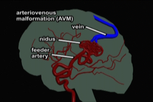
In an arteriovenous malformation, this normal branching structure is absent. Instead, we see a large feeder artery entering a nest (nidus) of small abnormal arterioles.
On the venous side, the normal branching structure is also absent, with one large vein draining the nidus.
Here is a view of the AVM as seen on angiogram.
The lack of a normal branching structure in the AVM results in extremely high flows and pressures across the abnormally small vessels of the nidus. As a consequence, AVMs have a high potential for rupture and hemorrhage of nidus vessels.
Intracerebral Hemorrhage
Hematoma
As mentioned previously, intracerebral hemorrhage takes place within the substance of the brain tissue itself. The accumulation of blood, also known as a hematoma continues to grow until the pressure surrounding it limits expansion or until it decompresses itself by rupturing into the cerebral spinal fluid system, either into the ventricles of the brain, or onto the surface of the brain. As seen here on an MRI image, the size of the hematoma can be very significant.
Neuronal Damage
Intracerebral hemorrhage can have a devastating effect on brain function in affected individuals. The hemorrhage occurs within the brain substance where billions of neurons are densely packed together. As the hematoma grows, it twists and ruptures neurons along its path causing massive irreversible damage. One neurosurgeon has likened the process to “turning on a fire hose in a bowl of jello”.
Switching from the microscopic to the macroscopic level, we see how the hematoma fragments brain tissue as it continues to expand. As a result of the very large amount of brain tissue destroyed in this process, intracerebral hemorrhage often has a catastrophic effect on the neurological functioning of affected individuals.
Cal Shipley, M.D. copyright 2020

