Carpal Tunnel Surgery Transcript
Carpal Tunnel Surgery
This is Dr. Cal Shipley with a review of surgery to treat Carpal Tunnel Syndrome.
For detailed information on carpal tunnel anatomy as well as the causes and pathophysiology of carpal tunnel syndrome, please see my video on carpal tunnel syndrome located in the Neurology Library of my website at trialimage.com.
Skin Incision
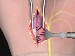
The surgery begins with a skin incision of several centimeters, starting at the wrist crease and running along the medial border of the thenar eminence.
Using these landmarks ensures that the incision is at safe distance from the median nerve.
Exposure of the Distal Flexor Retinaculum and Transverse Carpal Ligament
Blunt dissection and a self-retaining retractor are used to expose the distal flexor retinaculum and the transverse carpal ligament. Microsurgical scissors are used to open the distal flexor retinaculum and transverse carpal ligament. A retractor is used to visualize the remaining portion of the DFR.
The DFR extends distally to the superficial palmar arch artery. A scalpel is used to open the DFR up to the palmar arch.
Location and Separation of the of the Median Nerve
A Freer elevator is used to ascertain the precise location of the median nerve. The Freer elevator is then used to separate the median nerve from the transverse carpal ligament. The Freer elevator is then reinserted to protect the median nerve so that the remaining portion of the transverse carpal ligament and a portion of the antebrachial fascia can be opened.
Opening of the Antebrachial Fascia
Microsurgical scissors are again employed to open up the remainder of the TCL and the distal portion of the antebrachial fascia. The distal flexor retinaculum, the transverse carpal ligament, and a portion of the antebrachial fascia have now been opened. As a result, pressure within the carpal tunnel has been reduced and there is significant decompression of the median nerve.
Detachment of Adhesions
Depending on the case, as a result of inflammatory processes within the carpal tunnel, there may be extensive scar tissue adhesions attached to the median nerve. If these adhesions are not excised from the nerve during surgery, traction on the nerve with normal day-to-day use of the hand and wrist, they cause further injury and symptoms. Once these adhesions have been detached, the decompression of the median nerve is complete.
copyright 2023 Cal Shipley, M.D.

