Circle of Willis and Aneurysms Transcript
Circle of Willis and Aneurysms
This is Dr. Cal Shipley with a review of the circle of Willis, berry aneurysms, and subarachnoid hemorrhage in the brain.
Circle of Willis Anatomy
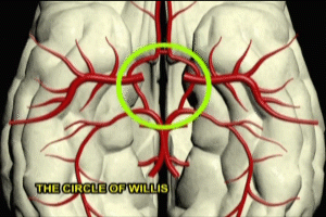
To begin, let’s take a look at the anatomy of the circle of Willis. The circle of Willis is best seen by examining the underside of the brain. The circle of Willis is really a beautiful piece of anatomy and represents a central hub from which radiates the entire blood flow to the brain.
Inflow is primarily via the internal carotids and vertebral arteries.
Outflow of blood from the circle occurs via anterior middle and posterior cerebral arteries, each with left and right branches on either side of the circle. The basilar, posterior communicating and anterior communicating arteries complete the circle.
By merging the inflow of blood to the brain from the carotid and vertebral arteries, the chances are minimized that an obstruction or impairment in flow in any one of these arteries will result in a significant impairment of blood supply to the outflowing arteries.
Berry Aneurysms and Subarachnoid hemorrhage
Now that we have a better understanding of the anatomy of the circle of Willis, let’s take a look at how it contributes to subarachnoid hemorrhage in the brain.
Aneurysms in the Circle of Willis
While beautiful in design and function, the circle of Willis is also the location of 85% of all aneurysms within the brain. One common site for the occurrence of an aneurysm is the junction between the anterior communicating and anterior cerebral arteries.
Aneurysm Formation
Aneurysms form as a result of an acquired structural weakness in the blood vessel wall. Arteries which carry very high flows, such as those in the circle of Willis, are thought to be particularly susceptible to mechanical damage in the vessel wall. Contributory factors such as cigarette smoking or high blood pressure may accelerate the process of damage to the vessel wall.
The strength of the aneurysm wall is inversely proportional to its size. Lesions one centimeter or larger are particularly susceptible to a sudden increase in growth and rupture.
Aneurysm Rupture
With rupture, blood spreads rapidly across the surface of the brain via the cerebrospinal fluid. Bleeding generally only lasts a few seconds. However, rebleeding may occur and is most likely within 24 hours of rupture and initial hemorrhage.
Morbidity and Mortality after Aneurysm Rupture
The most common cause of death and disability after subarachnoid hemorrhage is vasospasm: extreme narrowing of the arteries with impairment of blood flow to brain tissue and subsequent stroke.
Vasospasm usually occurs within the first three days after hemorrhage and generally peaks in effect about one week out. Vasospasm occurs as a result of chemical irritation of blood vessel walls by breakdown products of hemorrhaged blood. The duration of vasospasm and the location of the vessels affected determines the size and depth of subsequent strokes.
Cal Shipley, M.D. copyright 2021

