Fungal Meningitis Transcript
Fungal Meningitis
This is Dr. Cal Shipley with a review of fungal meningitis related to a contaminated epidural steroid injection.
In September 2012, the Center for Disease Control began an investigation into a multi-state outbreak of fungal meningitis related to epidural injection of contaminated steroids produced by the New England Compounding Center, a company based in Framingham, Massachusetts. Ultimately, the CDC investigation determined that patients in 20 states were involved in the outbreak, with 751 cases of meningitis and 64 deaths.
Spinal Cord Anatomy
To better understand the pathophysiology of these cases, let’s review some anatomy first. Rotating the spine to the side or lateral view, you zoom in for a close up of the second and third lumbar vertebrae. In most individuals, the spinal cord proper ends around the level of the second or third lumbar vertebra. A bundle of nerve roots known as the cauda equina originate from the end of the spinal cord and supply nerves to the lower lumbar and sacral segments.
Epidural Space
Surrounding the cauda equina is the epidural space. The epidural space at the level of the cauda equina is considered a preferable area for an injection, whether for injection of steroids or anesthetic, as the spinal cord above this level takes up more space and is more liable to injury.
Seen here in blue, the epidural space extends all the way up to the top of the vertebral column. On the left side of the screen is a view from above (cranial view) of a vertebra in cross-section, showing the relationship of the spinal cord and the epidural space.
Epidural Steroid Injections
Tens of thousands of epidural steroid injections are performed in the United States each year. They are primarily used in patients with nerve root impingement as a result of a herniated lumbosacral disc, also known as radiculopathy; spinal stenosis or narrowing of the spinal canal with impingement on the spinal cord, and nonspecific low back pain.
Despite the large numbers of procedures performed each year, the data is mixed in terms of effectiveness, with the most consistent benefits seen for short term relief of radiculopathy.
New England Compounding Center Case
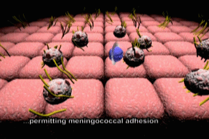
In the NECC fungal meningitis cases, several lots of methylprednisolone were found to be contaminated with fungal organisms.
These organisms were then injected into the epidural space, along with the methylprednisolone. The fungus spread throughout the epidural space, eventually reaching the brain.
Slow Growth of Fungus
Compared to bacteria, fungal organisms reproduce at a very slow rate. As a result of this characteristic slow growth, symptoms took several months to occur in many of the affected patients. Eventually, the fungal infection caused meningitis, inflammation of the meninges (coverings) of the brain.
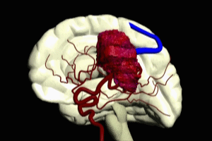
Fungal Meningitis, Vasculitis and Stroke
Many of the patients affected in the outbreak had injuries to their brainstem. The brainstem plays a critical role in the regulation of essential bodily functions, such as respiration. The inflammation of the meninges caused the adjacent arterial structures to become inflamed as well, a condition known as vasculitis. This may cause narrowing of the arteries, which results in the slowing of blood flow and the promotion of clot formation.
The formation of a clot, also known as a thrombus, will stop blood flow to that portion of the brain supplied by the inflamed artery. This may cause death of brain tissue, also known as a stroke. The severity of the clinical symptoms is directly related to the amount of brainstem tissue affected by the stroke, and can be fatal.
Vasculitis may also result in hemorrhage from the affected artery. The inflammation may cause a progressive loosening of the bonds between the cells in the artery wall and can lead to rupture of the artery. Once the artery has ruptured, blood hemorrhages into the brain tissue.
The rupture of arteries affected by vasculitis can cause a hemorrhagic stroke. As in clot-based stroke, in hemorrhagic stroke, rupture of the artery interrupts blood flow to brain tissue. Unlike clot-based stroke, the blood products that seep into brain tissues after artery rupture can cause further damage to the brain, often in a delayed onset fashion.
Please feel free to browse the links below the viewing screen on this page for more detailed information on many of the concepts touched on.
Cal Shipley, M.D. copyright 2020