Myocardial Infarction and Stroke CVS027
Myocardial Infarction and Stroke Transcript
Myocardial Infarction and Stroke
This is Dr. Cal Shipley with a review of embolization to the brain as a result of impaired left ventricular function.
Myocardial Infarction and Congestive Heart Failure
Two common conditions of the heart which can result in clot formation in the left ventricle and, then embolization of the clot to the brain, are congestive heart failure and myocardial infarction, (also known as heart attack). The physiological mechanism which gives rise to the clot formation is similar in both conditions.
Left Ventricular Function and Ejection Fraction
Let’s take a look at our heart model to better understand how this occurs. Taking a look at the heart in cross-section, we can isolate the left ventricle and then freeze it at its point of maximum contraction. The percentage of the blood which is contained within the left ventricle and which is ejected with each contraction is known as the ejection fraction. A normal typical ejection fraction is 50% to 65%. In other words, with normal contraction, 50% to 65% of the blood contained in the left ventricle just prior to contraction is ejected into the aorta.
Blood Stasis and Thrombus (Clot) Formation
In both myocardial infarction, (heart attack), and congestive heart failure, there may be marked impairment of left ventricular contraction, and therefore a much-reduced ejection fraction. This impairment of contraction can lead to a condition known as blood stasis. Stasis refers to pooling of blood in the base, (also known as the apex) of the left ventricle.
Embolization of Thrombus
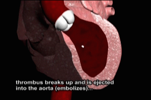
When blood pools anywhere in the human body, and the left ventricle is no exception, it tends to form clots. The medical term for a clot is a thrombus. Immediately after formation, a thrombus may be adherent to the wall of the left ventricle, and unpredictably, they may break free and be ejected into the aorta. The thrombus has now become an embolus.
Many thrombi have a tendency to break up into smaller pieces. The tiniest fragments may not cause significant obstruction to blood flow within the brain, however, larger pieces may cause significant interruption of blood flow to brain tissue, and subsequent stroke.
CT Imaging of Stroke
In this case, a significantly sized thrombus has lodged in one of the branches of the right middle cerebral artery. On CT scan, the area of stroke can be easily seen as this dark oval area. This particular patient had had a previous stroke, which is pointed out here on the right side of the screen.
Cal Shipley, M.D. copyright 2020

