Cataract Surgery Transcript
Cataract Surgery: Incidence
This is Dr. Cal Shipley with a review of cataract surgery. Lens cataracts are the most common cause of blindness worldwide, with up to 15 million people affected at any given moment. Cataract surgeries in the United States are among the most commonly performed, with three million procedures annually.
The Crystalline Lens
The structure of the human lens, also known as the crystalline lens, is a highly ordered and complex arrangement of specialized cells containing a high content of cytoplasmic proteins. These proteins, known as crystallins, impart the characteristic transparent quality to the lens.
Unlike other epithelial tissues in the human body, the crystalline lens does not shed non-viable cells. As a result, it is much more susceptible to the degenerative effects of aging.
Cataract Causes
In the industrialized world, age has clearly been demonstrated to be the greatest risk factor for formation of cataracts. A dose-response relationship has been demonstrated between exposure to ultraviolet B light waves found in sunlight and formation of lens cataracts. A relationship has also been shown to exist between smoking cigarettes and formation of lens cataracts.
It has been suggested that up to 20% of cataracts in the United States are related to smoking. As of this recording, hard scientific data implicating other risk factors is hard to come by. Many researchers, however, feel that one or more of the following factors may be contributory: diabetes, alcohol consumption, malnutrition, metabolic syndrome, and systemic corticosteroid use.
In addition, some researchers believe that daily use of inhaled high-dose corticosteroids, such as used by asthmatics, and also long-term use of statins, used to lower lipids, may increase the chance of cataract formation.
Normal versus Cataract Lens
Here is a comparison of a normal crystalline lens on the left and a lens affected by dense cataract formation on the right. The appearance of a cataract may vary considerably in terms of color and apparent texture. The one demonstrated here is a fairly common type.
As is apparent, the lens affected by the cataract has a much-reduced ability to transmit light information to the retina. Shown here is a comparison between vision with a normal lens, and vision with the type of dense cataract depicted in the previous image. Not only is there a dramatic reduction in the clarity of object perception, but color vision is also lost.
Cataract Surgery Techniques
There are primarily two surgical techniques used to remove cataracts in the United States.
Extracapsular Cataract Extraction
The oldest technique, also known as the standard technique, is called extracapsular cataract extraction. Also known as ECCE.
With this technique, the whole lens is removed at once. This requires a larger incision, and generally also requires sutures to close the incision. Suture reaction may cause astigmatism postoperatively.
Phacoemulsification
The second technique, now the most widely used in the United States, is called phacoemulsification. In this technique, the lens is removed in multiple pieces. This requires a smaller initial incision and generally no sutures.
The cataract surgery technique that I’m going to demonstrate is phacoemulsification, which has become the most popular approach to cataract removal in the United States. I should also preface this presentation by saying that although there are specific variations from surgeon to surgeon, the essential elements of phacoemulsification will remain the same. In addition, if you’re interested in learning more about the anatomy of the eye, I have created a companion piece to the cataract surgery segment, “Eye Anatomy Cataract Surgery”. This video is also in the Ophthalmology Library.
Anesthesia for Cataract Surgery
Just a quick review of anesthesia for cataract surgery. Anesthetic may be topical – simply by applying a liquid anesthetic to the outer surface of the eye without injection; peribulbar anesthesia, where anesthetic is injected above and below the globe of the eye; and retrobulbar anesthesia, where the anesthetic is injected behind the eye. The choice of which anesthetic to employ is dependent on patient comfort, and the preference of the surgeon performing the procedure. Topical anesthesia carries with it the least risk of complications, but may not provide adequate relief for patients experiencing discomfort with the lighting setup and manipulations of the eye required during the surgery.
Peribubar and retrobulbar anesthesia, on the other hand, provide a deeper level of anesthetic for most patients, but also carry with them the risk of complications, such as puncture of the globe, or of major arterial structures. Once the anesthetic has been administered, a pupillary dilating medication is applied externally to the eye, and retractors are placed to move the eyelids away from the operative field.
Cataract Surgery by Phacoemulsification
Peritomy Incision
To start the procedure, a small superficial incision is made in the conjunctival membrane at the edge of the cornea. This incision may theoretically be placed at any point along the circumference of the corneal edge, but in practice, the location depicted here is fairly common. Once the conjunctival incision has been made, a cutting tool is then used to extend this incision through the cornea and into the anterior chamber. This incision is known as a limited peritomy, and provides access to the lens.
This side view of the eye in cross-section shows how the peritomy incision provides access to the anterior chamber, and hence, to the lens. One of the key advantages of the phacoemulsification technique when used to remove a cataract is that this peritomy incision is much smaller than used in the standard ECCE technique previously mentioned. A smaller peritomy incision is then created to allow access for a second instrument during the procedure. A gel-like substance known as viscoelastic is then injected into the anterior chamber.
The viscoelastic prevents the cornea from collapsing and maintains the volume of the anterior chamber, providing the surgeon with adequate room to manipulate instruments during the procedure. The lens is completely contained in a membranous envelope known as the lens capsule. The final step in gaining direct access to the lens is to create an opening in the lens capsule. This is known as a capsulotomy. The tapered tip of an instrument is used to create the opening.
Hydrodelineation and Hydrodissection
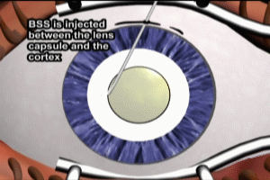
Once the circumferential incision is complete, the loose flap is removed. The lens is now accessible. A side view in cross-section shows the capsulotomy with a portion of the front or anterior aspect of the lens capsule removed. As seen in a cross-section here, the lens consists of three layers. An inner nucleus surrounded by two thinner layers, the epinucleus, and the cortex. In order to protect the lens capsule from injury during manipulation of the lens, it is important to separate the epinucleus and cortex from both the lens capsule and the nucleus of the lens. The process of separating the epinucleus from the nucleus of the lens is known as hydrodelineation.
A balanced salt solution is injected between the epinucleus and the nucleus, and hydrostatic static pressure separates the two layers as depicted here. The separation of the epinucleus from the nucleus of the lens allows the lens nucleus to be freely manipulated without undue stress on the capsule. As an added precaution, and in a similar fashion, the cortex is separated from the lens capsule. This process is known as hydrodissection. As with hydrodelineation, a balanced salt solution is injected between the cortex and the lens capsule.
The separation of the cortex and the lens capsule further reduces the chances for stress on the capsule during manipulation of the lens nucleus. Once hydrodelineation and hydrodissection have been completed, the lens nucleus may be freely moved without stressing the capsule. Preserving the structural integrity of capsule is critical, as once the cataract has been removed, the lens capsule must hold the intraocular lens implant. Now that the lens is accessible, the phacoemulsification process can proceed. A secondary instrument is inserted through the smaller peritomy incision to assist with stabilization and manipulation of the lens during emulsification process.
Phacoemulsification
The tip of the phacoemulsification instrument is then introduced through the primary peritomy incision. The tip of the phacoemulsification instrument consists of a steel or titanium needle with a hollow core. The tip vibrates at an ultrasonic frequency, which emulsifies, or breaks up, the cataract as it comes into contact with it. Fragments of cataract are then suctioned, or aspirated, through the hollow core. The phaco tip is used to create grooves in the lens nucleus.
Several passes are made and the groove deepens with progressive removal of cataract material. Care is taken to keep the ends of the grooves away from the outer edge of the cataract, so as to avoid injuring the lens capsule. As seen in this cross-section view, the depths of the grooves are kept to about 80% of the thickness of the cataract, another measure to prevent injury to the lens capsule.
The phaco tip is used to create additional grooves in the cataract, forming a pattern reminiscent of the 1960s peace sign. With the assistance of the secondary instrument, the cataract is rotated 180 degrees, and the grooving process repeated. Now that the grooving has been completed, a process known as cracking is utilized to remove the remainder of the cataract material in the grooves, and separate the cataract into several smaller pieces. Once the cracking has been completed, the cataract has been divided into six pieces. With the assistance of the secondary instrument, the phaco tip is used to aspirate the cataract fragments.
The cataract nucleus has now been removed. The epinucleus and cortex layers still remain within the lens capsule, and must be removed before implantation of the intraocular lens can be performed. A suction instrument is used to remove the layers. In the cross-section side view, the epinucleus, seen in red, is removed, followed by the cortex, depicted in blue. With the epinucleus and cortex layers removed, the lens capsule is now ready to accept the intraocular lens implant.
Intraocular Lenses (IOL)
There are many types of intraocular lenses, or IOLs, now available, for implantation after phacoemulsification removal of a cataract. Their key characteristic is that they must be foldable so that they can be introduced through the relatively small peritomy incision. For the purposes of this presentation, I am going to be using a foldable intraocular lens, known as a toric lens. Toric lenses are manufactured in one piece and are particularly useful for correcting astigmatism.
The intraocular lens implantation is relatively straightforward. The folded IOL is contained in a plastic tube, which is introduced through the peritomy incision. The IOL is then pushed out of the tube into the lens capsule, where it unfolds. An instrument may be used to assist in the unfolding process. Once the IOL has completely unfolded, the surgeon rotates it to a predetermined position, so that astigmatism will be properly corrected. The series of markings on the periphery of the IOL are used to rotate it to the correct position. The removal of the cataract and the implantation of the intraocular lens is now complete.
Cal Shipley, M.D. copyright 2020

