Eye Anatomy in Cataract Surgery Transcript
Eye Anatomy in Cataract Surgery
This is Dr. Cal Shipley with a review of the anatomy of the eye. The human eye is a beautifully complex and sensitive instrument for interpreting the light energy which is part of our world. Let’s take a look at how it’s put together.
Eye Anatomy: the Globe
The globe of the eye is a spherically shaped structure which is hollow, and consists of three layers. Seen here in cross section, they are: the sclera, the white outer portion of the globe; the middle layer, or choroid, which carries most of the blood supply to the inner eye; and the inner, light sensing layer, the retina. The retina is composed of several layers of light sensitive neurons which come together at the back of the globe to become the optic nerve.
Cornea
Projecting from the front, or anterior portion of the globe, and occupying about one sixth of the total globe diameter, is the transparent cornea. The cornea provides a protective covering over the iris, pupil, and anterior chamber of the eye. The cornea refracts light, and is responsible for about 65% of the focusing power of the human eye. The curvature of the cornea is fixed, and therefore, so is its focus, whereas, the curvature of the eye lens may be adjusted to compensate for an object’s distance. We’ll see how this variable focus mechanism works in just a moment.
Iris
The iris is a thin circular structure which gives the eye its characteristic color, and whose primary functional units are muscle cells. Its inner border forms the pupil and a set of muscles, the dilator papillae, contract to enlarge the pupil, while another set of muscles, the sphincter pupillae contract to reduce pupillary size. Thus the iris’s primary function is to control the amount of light entering the eye. The iris also constricts to decrease pupillary size when re-focusing on an object close to the eye. This is known as the accommodation reflex. It is controversial as to whether or not the decrease in pupillary size assists with focusing during the accommodation reflex.
Lens Capsule
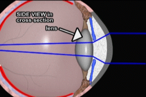
Lying directly behind or posterior to the iris is the lens capsule which contains the lens. Attached to the outer rim or equator of the lens capsule are a series of fibers known as zonules. At the other end, the zonules are attached to the ciliary body. The zonules act as suspensary ligaments for the lens, and keep the lens under tension in the resting state.
Ciliary Body
Switching back to the front view, if we cut the globe back a little bit further, we reveal the ciliary body attached to the zonules. To change the curvature and enhance the focus of the lens, the muscles of the ciliary body contract inward in a radial fashion. This in turn permits the lens to expand, increasing its curvature and changing its focus. To change the focus to objects further away, the ciliary body relaxes. This re-establishes the pull of the zonules on the lens, flattening its curvature and permitting focus on more distant objects.
Lens
Let’s take a closer look now at the structure of the lens, also known as the crystalline lens. The lens is contained within the lens capsule, and is a multilayered structure. Seen in cross section, the innermost component of the lens is the nucleus. The nucleus is surrounded by two, thinner outer layers, the epinucleus and the cortex. The outermost layer is the lens capsule. The capsule is by far the thinnest of the layers, averaging just several microns in thickness.
Eye Anatomy: Chambers
Now let’s take a look at the chambers and vitreous of the eye. As previously mentioned, the anterior chamber lies between the cornea and the iris. The posterior chamber is situated in the space created by the iris, the ciliary body and the zonules as shown here.
Aqueous Humor
A watery substance, the aqueous humor flows throughout both anterior and posterior chambers. The aqueous is produced by the ciliary bodies, circulates throughout the posterior and anterior chambers, and then is re-absorbed in the anterior chamber through the trabecular meshwork, located at the peripheral margin of the iris, where it inserts into the ciliary body.
The aqueous humor provides nutrients to the cornea and the lens, neither of which have their own blood supply. The aqueous humor also provides inflationary pressure in the anterior chamber to maintain proper expansion and curvature of the cornea. A condition known as glaucoma occurs when intraocular pressures are too high, usually as a result of inadequate re-absorption of aqueous humor.
Vitreous Humor
The vitreous humor, or vitreous, is a gell-like substance which fills the posterior two thirds of the globe. The vitreous is bordered in front by the lens, the zonules and the ciliary bodies, at the back by the optic nerve and the retina, and on all sides by the retina. Like the aqueous humor, the vitreous is produced by the ciliary bodies. Unlike the aqueous humor, the vitreous does not circulate and remains more or less static throughout life.
Like the aqueous humor in the anterior chamber of the eye, the vitreous serves to maintain the shape of the globe by exerting outward pressure. This pressure helps to keep the retina in close contact with the choroid plexus, which supplies the retina with oxygen and nutrients from the blood stream. Despite the fact that the vitreous is in contact with virtually 100% of the retina, it is only attached at the optic disc, and where the retina ends anteriorly, the ora serrata.
Eye Anatomy: Retina and Optic Nerve
Finally, let’s turn our attention again to the retina. You may notice that the top, or cranial, view of the eye in cross-section looks very similar to the side view. This is because the geometry of the globe and the cornea are based on symmetrical spheres, as are the retina and the choroid layer. In addition, the geometry of the lens is based on an almost symmetrical circular disc with the zonules and ciliary bodies arrayed in a circular, radial fashion around it.
The retina, pictured in red here, lies against the choroid layer and between the choroid and the vitreous. As previously mentioned, the choroid plexus provides the blood supply to the retina. The vitreous helps to keep the retina in place.
Viewing the anterior of the eyeball from a frontal view, you see the tissue of the retina occupying the globe from top to bottom and from left to right. The disc of the optic nerve is visible, with branches of the retinal artery radiating from it. The centrally located macula is also present, and the center of the macula is the fovea. The fovea contains the highest concentration of cone photo receptors in the retina, and is responsible for sharp central vision and color discrimination.
Returning to our cranial view of the left eye in cross section, we see the optic nerve in pale yellow, the retinal artery, and the area of the macula and foveal pit. The area of the macula containing the fovea receives light from the center of the visual field of view, and provides the greatest acuity of vision in the human eye.
Cal Shipley, M.D. copyright 2020

