Lumbar Disc Decompression
This is Dr. Cal Shipley with a review of decompression of a bulging disc. As an example, today we’re going to take a look at a bulging intervertebral disc between the fourth and fifth lumbar vertebra in the low back.
Anatomy
Taking a look first at the anatomy, we see the fourth and fifth lumbar vertebrae, or L4 and L5 as they’re known for short, and the first sacral vertebra, or S1, and then the L4-L5 disc sitting between the fourth and fifth lumbar vertebra, and the L5-S1 disc sitting between the fifth lumbar and first sacral vertebrae.
The intervertebral discs are made of a firm but flexible material that act as shock absorbers between the spinal vertebrae. Situated as they are at the bottom of the spine, the L4, L5 and S1 vertebrae, and their accompanying discs, bear considerable stress as a result of the weight and movement of the upper body, and as a result, have a greater frequency of injury and degenerative change on their counterparts higher up in the spine (the exception to this rule being the vertebrae of the neck).
Spinal Nerves and Spinal Cord
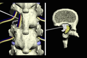
Finally, we see the spinal nerve roots exiting to the left and right between the vertebrae.
Rotating to a view from above of L5, the anatomical relationships of all these structures become a little more readily apparent. The spinal cord ends at the level of the second lumbar vertebra, giving off multiple branches which become the nerve roots to the remaining lumbar and sacral vertebral segments. These lower nerve roots when bundled together in the spinal canal are known as the cauda equina. The left and right spinal nerve roots branch off of the cauda equina and the intervertebral disc sits on top of the vertebra, and therefore between the L5 and L4 vertebrae.
Disc Bulge
Okay, let’s zoom in a little bit on the anatomy here and take a closer look at how a disc bulge comes about. As mentioned previously, the discs between the fourth and fifth lumbar, and fifth lumbar and first sacral, verterbrae are particularly susceptible to injury. Depending on the severity and direction of the bulge, the spinal nerve roots or the nerves of the cauda equina may be compressed resulting in numbness, pain or weakness in the tissues and muscles supplied by the compressed nerves.
Disc Structure
An examination of the inner structure of the disc makes it easier to see how age related degeneration or trauma may result in disc bulge. Seen in cross section, the disc is composed of two primary structures, the annulus, a tough fibrous outer shell, and the nucleus, the softer inner core. The annulus gives the disk its strength, while the nucleus provides flexibility and cushioning.
Disc Herniation
Either as a result of age-related degenerative change or trauma, tears may occur in the annulus. These tears weaken the annulus, resulting in a loss of its structural integrity and leading to bulging of the disk as a result of the pressure exerted from the spine above.
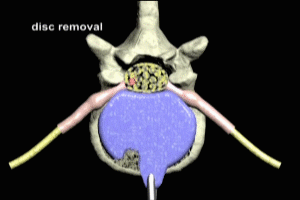
Weakening of the annulus fibrosis may ultimately result in through and through tears of its fibers with extrusion of the nucleus pulposus outside the confines of the annulus as seen here. This is known as a disc herniation. This example shows a pretty extreme herniation with severe compression of the spinal cord.
Symptomatic disc herniations generally require aggressive surgical treatment with complete removal of all disc material.
Symptomatic bulging discs, however, may be treated more conservatively with a technique known as coblation and coagulation.
Lumbar Disc Decompression with Coblation
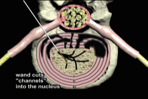
Utilizing a radiological technique known as fluoroscopy, the coblation wand is guided through the back of the patient and inserted into the nucleus of the affected disc. An electrical charge is then applied to the coblation wand, which is then advanced into the nucleus, vaporizing nuclear material as it goes and cutting a channel. This vaporizing and cutting action is known as coblation.
With repeated insertion and withdrawal, multiple channels are cut into the nucleus.
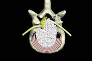
Each time the coblation wand is withdrawn, the electrical charge applied to it is changed resulting in coagulation of the tissue on either side of the cut channels. The coagulation pulls the walls of the channels together, resulting in an overall shrinkage of the size of the nucleus and therefore of the disc.
When successful, coblation and coagulation can reduce overall disk size by 10 to 15% resulting in a reduction in the disc bulge. Many patients have a marked improvement in symptoms as a result. As mentioned previously, this technique is not effective for ruptured or herniated discs.
Cal Shipley M.D., copyright 2020