Saddle Pulmonary Embolism PUL044
Saddle Pulmonary Embolism Transcript
Saddle Pulmonary Embolism
This is Dr. Cal Shipley with a review of Saddle Pulmonary Embolism.
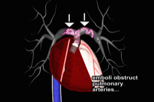
The right and left pulmonary arteries arise from a common root in the right ventricle. They receive deoxygenated blood from the right ventricle and then carry blood through the lungs to be re-oxygenated. Any significant impairment of blood flow through the pulmonary arteries can have catastrophic effects. The area where the left and right pulmonary arteries bifurcate, or split off, from the main pulmonary route is also known as the pulmonary artery saddle.
Here we see a deep venous thrombosis (or DVT) in a deep vein of the left lower limb. If the DVT becomes dislodged, it may begin to travel through the venous system. It is now termed an embolus. It is not uncommon for larger emboli to fragment as they move through the venous system.
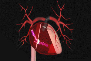
Ultimately, the emboli reach the heart where they enter through the right atrium, pass through into the right ventricle, and then are impacted into the root of the pulmonary artery by contractions of the right ventricle.
Trapped in the pulmonary artery saddle, the emboli may cause blood flow through the arteries to come to a virtual standstill. Severe impairment of blood flow through the pulmonary arteries results in a drastic reduction in oxygenation of all bodily tissues and organs. If this situation cannot be corrected rapidly, loss of brain and cardiac function will result in death.
The primary difficulty in treating patients affected by Saddle Pulmonary Embolism is that this whole process may occur in just a few minutes, leaving very little time for effective resuscitation efforts.
Cal Shipley, M.D. copyright 2020


