Throat Anatomy and Endotracheal Intubation RES006
Throat Anatomy and Endotracheal Intubation Transcript
Throat Anatomy and Endotracheal Intubation
This is Dr. Cal Shipley with a review of throat anatomy and endotracheal intubation.
Anatomy of the Throat
Musculature
The musculature of the neck and throat is highlighted by the sternocleidomastoid muscles ,both of which have two heads which attache to the sternum and clavicle on either side.
Airway
Removal of the muscular layer reveals the trachea, the major airway connecting the nose and mouth to the lungs and the cricoid cartilage, which provides an attachment point for multiple muscles and ligaments involved in the production of speech and opening and closing of the airway.
Cervical Spine
Deep to the trachea lies the cervical spine. The cervical spine literally acts as the backbone of the throat and neck providing major structural support as well as providing a protected conduit for the spinal cord.
Vocal Apparatus
On closer examination, we see the thyroid and cricoid cartilages and housed within the thyroid cartilage, the vocal cords.
Pharynx, Larynx and Epiglottis
Switching to a side view, we see the root of the tongue, the trachea again ringed by its external cartilages and behind the trachea, the esophagus. A cross-sectional view reveals the epiglottis, a flap of fibrocartilaginous tissue which is attached anteriorly to the inner aspect of the thyroid cartilage. Lying posteriorly at the very back of the throat is the cervical spine.
It is worth noting here that the area bordered superiorly by the epiglottis and inferiorly by the cricoid cartilage and enclosing the vocal apparatus is known as the larynx.
The pharynx refers to that area of the airway lying directly above the vocal chords and the epiglottis. The purpose of the epiglottis is to allow for free movement of air in and out of the trachea during breathing and to close over the airway during swallowing to force oral secretions and food down the esophagus and prevent their movement into the lungs.
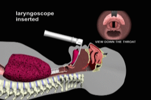
The inset on the left depicts a view looking down the throat as one might see it during endotracheal intubation. It is critically important to a safe and successful intubation to the operator be very familiar with the anatomical landmarks noted here. Again, we can observe the closure of the epiglottis during swallowing.
Endotracheal Intubation
Now that we’ve set up the relevant anatomy, let’s turn our attention to endotracheal intubation. Endotracheal intubation is used whenever an assured protected airway is necessary in a patient, such as in emergency situations where the patient may be unconscious or have impaired spontaneous respirations or during general anesthesia. A protected airway refers to the fact that the airway is protected from aspiration into the lungs of oral contents.
We’ll see in just a few moments here how a properly inserted endotracheal tube protects against aspiration.
In a cross-sectional view, with the patient prone, the key anatomical landmarks are noted. A laryngoscope is used to accomplish the intubation. The laryngoscope has a removable metal blade. Laryngoscope blades come in a variety of sizes and shapes to accommodate variations in the throat and neck anatomy from patient to patient.
Endotracheal Intubation Technique
Prior to insertion of the laryngoscope, the head of the patient is extended slightly on the cervical spine, pushing the chin upward and reducing the angle between the oropharynx and the upper airway.
Blade Insertion
The blade of the laryngoscope is inserted to depress the tongue and open the epiglottis. It is critical that the blade chosen be of an appropriate shape and size for a given patient so that the tip of the blade can reach the hyoepiglottic ligament at the base of the epiglottis.
When pressure is applied to the ligament as shown here, the epiglottis opens fully.
Tube Insertion
The endotracheal tube is now inserted. Typically, a length of heavy but flexible wire is inserted through the endotracheal tube to conform to the shape of the airway and assist the operator in insertion.
The endotracheal tube is inserted so that the inflatable cuff is pushed beyond the vocal cords. The laryngoscope is now withdrawn. The cuff on the laryngoscope lies beyond the vocal cords in the upper trachea as noted here.
When the cuff is inflated with air, it anchors the endotracheal tube within the trachea and also seals the trachea against the possibility of aspiration. As mentioned previously, this is what is meant by a protected airway.
The patient is now ready for ventilation with either an Ambu bag or a ventilator machine. Once ventilation has begun, the lungs are auscultated to ensure airflow and eliminate the possibility of an esophageal intubation. Please see my animation on esophageal intubation in the Resuscitation Library for more information.
Cal Shipley, M.D. copyright 2021

