Aortofemoral Bypass Graft Transcript
Aortofemoral Bypass Graft
This is Dr. Cal Shipley with a review of occlusive disease of the aorta and aortobifemoral graft procedure.
Anatomy
Let’s take a look at some relevant anatomy first. In the abdomen, the intestines, stomach, and liver are enclosed in a membrane known as the peritoneum. The peritoneum also contains the blood vessels through which blood flows to and from the abdominal organs.
While it is flexible enough to allow for normal peristaltic wave action in the intestines, the peritoneum anchors and stabilizes both the intestines and the blood vessels and prevents them from twisting or kinking.
The peritoneum consists of front and back or anterior and posterior layers, and so, in effect, the organs which are contained within it are sandwiched between these two layers. Removal of the anterior layer reveals the small intestine and the colon or large intestine with its cecum, which connects to the small intestine, and from which originates the appendix.
We can now also see the back or posterior layer of the peritoneum. Organs and structures which lie behind this layer are known as retroperitoneal. Removal of the posterior layer of the peritoneum reveals the aorta and femoral arteries. The aorta lies behind the peritoneum, and therefore its location is termed retroperitoneal. The iliac arteries are situated between the aorta and the femoral arteries.
The Aorta
Originating from the left ventricle of the heart, the aorta carries oxygenated blood to all organs and tissues in the human body. The femoral arteries carry blood from the aorta to the lower limbs. Significant buildup of atherosclerotic plaque in the aorta above the level of the femoral arteries will cause severe impairment of blood flow to the lower limbs. Depicted here is a total obstruction of the aorta due to plaque buildup.
Aortofemoral Occlusive Disease
While the lower limbs may continue to receive a small amount of arterial blood flow via collateral vessels originating from aorta above the level of the obstruction, the severe decrease in overall blood flow requires urgent intervention to prevent loss of limb due to gangrene. At any given time, eight to 10 million Americans are affected by occlusive disease of the arteries. One form of which is aortic obstruction, as depicted here.
Symptoms of impaired arterial blood flow to the lower limbs, also known as ischemia, may consist of loss of hair on the affected limb, intermittent or chronic blue discoloration of the toes or feet (also known as cyanosis), and limb pain associated with minimal exercise, also known as intermittent claudication.
As obstruction to arterial blood flow progresses, pain may occur at rest, particularly over the heads of the metatarsal of the feet, and death of soft tissue around the toes and feet, also known as necrosis, may begin to occur as a precursor to gangrene.
Treatment of Aortofemoral Occlusive Disease: Angioplasty
Turning now to the treatment options for aortofemoral occlusive disease, and this includes obstruction due to plaque anywhere from the distal aorta, where we have depicted it in this presentation, to the iliac arteries, which lie between the femoral arteries and the aorta, or in the femoral arteries themselves. The first treatment option to look at is angioplasty. Angioplasty, for those of you who are not familiar, involves sliding a catheter into the obstructed artery and inflating a balloon which then compresses the plaque, and opens up the lumen of the artery.
Please go over to the cardiovascular library and look under angioplasty if you’re interested in seeing specifically how this works. The same technique that’s used in coronary arteries is also used in peripheral arteries, and can be used in aortofemoral occlusive disease. The main advantage of angioplasty is that it is less invasive than a surgical procedure and has a significantly lower rate of complications. The main disadvantage of angioplasty is the reoccurrence of obstruction in the treated arteries.
Unlike surgical bypass procedures, where the blood flow is rerouted around the diseased portion of the artery, with treatment by angioplasty, blood must flow through the diseased section of the artery. Reaccumulation of plaque in the diseased section of the artery may occur resulting in further obstruction to blood flow. Surgical treatment of occlusive aortofemoral disease represents the flip side of the treatment coin. Complication rates are higher and potentially more serious than with angioplasty treatment, but restoration of blood flow is better and longer-lasting.
In view of these advantages and disadvantages, current recommendations are that surgical treatment be used for patients with occlusive aortofemoral disease, with angioplasty being reserved for patients who have a life expectancy of less than two years or who have coexisting medical conditions, also called comorbidities, which preclude the safe use of surgical techniques.
Aortofemoral Bypass Graft
Let’s turn now to one surgical technique used in the treatment of occlusive disease of the distal aorta. A vertical incision is employed to open the abdomen. Once the layers of the abdominal wall and the anterior peritoneum are open, a series of retractors are used to move the intestines and expose the posterior layer of the peritoneum. The posterior peritoneum is then opened to expose the aorta.
Here, again, we see the area the distal aorta completely obstructed by atherosclerotic plaque. Here are the iliac arteries, which lie between the aorta and the femoral arteries.
Division of the Diseased Aorta
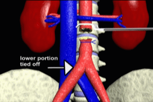
A vascular clamp is applied across the aorta above the level of obstruction. The diseased section of the aorta is then removed. The distal portion of the aorta is then tied off.
Dacron Graft
A synthetic Dacron graft is then stitched or sutured to the upper, or proximal, portion of the aorta.
While this particular technique I’m discussing today utilizes a synthetic graft, grafting may also be accomplished using veins which are harvested from the patient at the beginning of the procedure. The choice of a synthetic versus a venous graft is partly up to the personal preference of a surgeon, but may also be dictated by the presence or absence of good quality veins in a particular patient.
Also, in a situation where a bilateral bypass is required, a synthetic graft such as the one pictured here requires only three points of attachment, whereas patient veins would require four separate points of attachment, two points of attachment for each vein on either side of the aorta. Now that the upper portion (or proximal portion) of the graft has been attached, preparations must be made to attach the limbs.
Exposing the Femoral Artery
An incision is made in the groin to expose the femoral artery. Using two fingers of the hand, the surgeon gently separates the tissues in the retroperitoneal space. This is known as blunt dissection and avoids the use of instruments which may puncture or tear vital structures. This technique creates a retroperitoneal tunnel through which one limb of the Dacron graft will be pulled.
Positioning and Attachment of the Graft to the Femoral Arteries
A clamp, known as a Pean clamp, is used to pull the limb of the graph through the retroperitoneal tunnel. This clamp has a particularly blunt tip, which reduces the chance of injury to vital structures as it first grasps, and then pulls, the graft limb through the retroperitoneal tunnel. The creation of the retroperitoneal tunnel provides for an easier and safer passage of the Pean clamp. Vascular clamps are now applied across both limbs of the Dacron graft. The aortic clamp is removed. Blood now flows into the limbs under systemic arterial pressure. This serves two purposes. The previously attached portion of the Dacron graft is tested for leaks, and twisting or kinking of the limbs of the Dacron graft is prevented throughout the rest of the procedure. The Pean clamp is now inserted through the groin incision and into the retroperitoneal tunnel. It is inserted until the tip can be seen in the previously made peritoneal incision.
Here is a close up showing the relationship between the cecum of the large intestine, the iliac artery, and the tip of the Peon clamp as it moves safely through the previously created retroperitoneal tunnel.
Having safely navigated the retroperitoneal tunnel, the tip of the Pean clamp is opened, and the vascular clamp attached to the graft limb is used to bring the end of the limb towards the Pean clamp. The clamp is then closed to grasp the end of the limb and the vascular clamp removed.
The Pean clamp is then used to pull the graft limb through the retroperitoneal tunnel and into the groin incision. The left graft limb is pulled down into the area of the left femoral artery by the same technique.
Once both graft limbs have been sutured to their respective femoral arteries, the bypass procedure is complete. Normal blood flow to the lower limbs is restored.
Cal Shipley, M.D. copyright 2020

