Anatomy and Physiology of the Placenta OBS116
Anatomy and Physiology of the Placenta Transcript
Anatomy and Physiology of the Placenta
This is Dr. Cal Shipley with a review of the anatomy and physiology of the human placenta.
Placenta and the Gravid Uterus
For this presentation, I’m going to be looking at a uterus, which contains a fetus at about nine months of gestation. This is also known as a gravid uterus at term. The placenta is attached to the uterine wall and is connected to the fetus via the umbilical cord.
Placental Purpose
The primary function of the placenta is to facilitate transport of nutrients and waste products between the mother and the fetus.
Placental Anatomy
To better understand how the placenta works, let’s take a look at a view in cross section.
Placental Vascularity
Maternal uterine arteries known as spiral arteries supply blood to the placenta by penetrating the decidua layer. The maternal blood circulates through the intervillous space and then returns to the uterus via the endometrial veins. Branching structures known as the placental villi contain the fetal blood vessels.
The Chorion
The area of the placenta, which contains the villi is known as the chorion. The terms placental villi and chorionic villi are interchangeable. The outer borders of the villi are lined by trophoblast cells. The fetal blood vessels contained within the villi are branching structures of the umbilical arteries and the umbilical vein. The villi are bathed in maternal blood, but the trophoblast cell layer prevents any direct physical contact between the fetal and maternal bloodstreams.
Placental Physiology
The Trophoblast
Viewing one of the placental villi on a microscopic level we see the maternal and fetal blood separated by the trophoblast cell layer.
Throughout most of the period of fetal gestation, the trophoblast consists of two cell layers. The outer syncytiotrophoblast, which is in direct contact with the maternal blood, and the cytotrophoblast layer, which is in contact with the endothelium of the fetal capillaries (for purposes of graphics simplicity in this presentation, I have omitted the layer of capillary endothelium, which normally sits between the trophoblasts and the fetal blood.)
Oxygen Diffusion
As the gestation progresses, the cytotrophoblast atrophies, so that at full term there is just a single layer of cells separating the maternal blood and the fetal capillary endothelium. Oxygen diffuses across the trophoblast layer from the maternal blood to the fetal blood. This diffusion occurs because the pressure exerted by the oxygen molecules in the maternal blood, also known as the partial pressure of oxygen, is greater than the partial pressure of oxygen in the fetal blood.
Because the oxygen molecules do not require any additional chemicals substances to cross cell membranes, this process is known as passive diffusion.
Nutrient Transport
Nutrients, on the other hand, require biochemical assistance to cross the trophoblast cell layers.
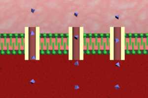
Let’s take a look at how the most important nutrient glucose traverses the cells of the trophoblast. Here’s a view under even greater magnification showing the maternal blood, the trophoblast cell wall, and within the cell wall, the cell cytoplasm.
The cell membrane consists of a double layer of lipids of a type known as phospholipids. Glucose molecules are water-soluble and will not dissolve in the presence of lipids. Because of this, glucose molecules depend on a special kind of protein known as an active transport protein in order to cross the trophoblast cell membrane. This process is known as facilitated diffusion. The active transport proteins allow the glucose molecule to both enter and then exit the trophoblast cell so that it can then cross the capillary endothelium and enter the fetal bloodstream.
A wide variety of essential fetal nutrients, such as amino acids, fatty acids, and iron also across the trophoblast cell layer with the help of transport proteins.
From villus capillaries, oxygen and nutrients are carried to the umbilical vein and through the umbilical cord to the fetal circulation. Oxygen and nutrients are distributed to the heart, brain, and all fetal organs and tissues.
Carbon Dioxide
Simultaneously carbon dioxide and other waste products flow from the fetus to the placenta via the umbilical arteries.
Compared to oxygen the situation with carbon dioxide in the maternal and fetal blood is reversed with the partial pressure of carbon dioxide in the fetal blood greater than that of the maternal blood. This sets up a pressure gradient, which causes the carbon dioxide to passively diffuse across the trophoblast cell layer and into the maternal blood.
The Haldane Effect
Diffusion of carbon dioxide across the placenta is also assisted by the Haldane effect. The Haldane effect describes the increased capacity of deoxygenated blood to carry carbon dioxide compared with oxygenated blood. As fetal blood absorbs oxygen to form oxyhemoglobin, it has reduced affinity for carbon dioxide and releases this carbon dioxide to the mother.
Waste Products
Waste products, such as urea, uric acid, and creatinine cross the placenta from the fetal to the maternal blood, largely as a result of concentration gradients. The principle involved in a concentration gradient is similar to the pressure gradients which result in passive diffusion of oxygen and carbon dioxide across the placenta.
When the concentration of waste products is higher in the fetal blood than in the maternal blood, passive diffusion of the waste products will always occur in the direction of the maternal blood.
Additional Functions of the Placenta
In addition to its primary role as a vehicle for the exchange of oxygen, nutrients, and waste products between the mother and the fetus, the placenta plays an important role in the synthesis of several hormones, which support fetal development and gestational success.
Here is a partial list of hormones synthesized by the human placenta. For the most part, the placenta relies on substrates supplied by either the fetus or the mother to achieve hormone synthesis.
Cal Shipley, M.D. copyright 2021

