Epidural Anesthesia
This is Dr. Cal Shipley with a review of epidural anesthesia.
Epidural Anesthesia Purpose
Epidural anesthesia is used to reduce pain sensation and is particularly useful in situations where it is desirable to have the patient conscious, such as during childbirth. To reduce pain during labor, a nerve block of the lumbar and sacral nerve roots coming off the spinal cord is necessary.
Cauda Equina
In most individuals, the spinal cord proper ends at around the level of the second lumbar vertebra. A bundle of nerve roots known as the cauda equina originate from the end of the spinal cord and supply of the nerves to the lower lumbar and sacral segments.
For safety’s sake, the needles and catheters for epidural anesthesia during childbirth are typically inserted below the level of the second lumbar vertebra in the area of the cauda equina in order to avoid the spinal cord. For purposes of our demonstration today, I have chosen the inter-space between the fourth and fifth lumbar vertebrae.
Epidural Anesthesia: Spinal Anatomy
Looking at the anatomy from both a view from above and a side view, we see the dural covering surrounding the cauda equina, and then inside of the dural covering, the arachnoid layer. The target of epidural anesthetic, the epidural space, lies between the dural covering and the rearmost portion of the spinal canal. Within the dura and arachnoid layers flows cerebrospinal fluid, or CSF, which originates from ventricles in the brain, not to be confused with the ventricles of the heart.
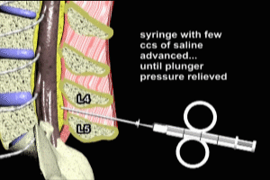
Procedure
Now that we’ve reviewed the relevant anatomy, let’s take a look at the procedure itself. For purposes of demonstration today we’re going to use the L4-L5 inter-space, but, in fact, any inter-space below the level of the second lumbar vertebra may be employed in an obstetrical epidural anesthetic. A long needle known as a Tuohy needle is first advanced into the soft tissues between the spinous processes of the fourth and fifth lumbar vertebra. A syringe containing a few CCs of saline is attached to the Tuohy needle. With the operator maintaining a light but steady pressure on the plunger of the syringe, the needle and syringe are advanced steadily through the inter-space. Once the tip of the needle is advanced into the epidural space, the operator notes a release of the syringe plunger as the saline squirts into the space. The epidural catheter is then inserted through the Tuohy needle. Owing to a slight curve at the tip of the Tuohy needle, the catheter is directed upward, approximately four to five centimeters into the epidural space. The Tuohy needle is then removed.
To ensure that the catheter is properly placed in the epidural space and not within the dura and arachnoid coverings, a syringe containing a small dose of anesthetic, typically lidocaine, is attached to the catheter and injected. A minimum of three minutes after the test dose, the patient’s ability to move their lower limb is tested. If the patient is able to move their lower limb, this indicates proper placement of the epidural catheter in the epidural space. If the lower limb cannot be moved, then placement of the catheter within the dura and arachnoid layers must be assumed. This is also known as an intrathecal catheter placement, and requires that the catheter be removed, and the placement procedure started over.
Once proper placement of the catheter has been ascertained, a bolus dose, consisting typically of about 10 CCs of anesthetic, is injected through the catheter to initiate the anesthesia. A continuous infusion of anesthetic is then initiated. The rate of the infusion may be adjusted according to the patient’s response and the level of anesthesia desired.
Cal Shipley, M.D. copyright 2020