Umbilical Cord Prolapse and Hypoxic Ischemic Encephalopathy OBS148
Umbilical Cord Prolapse and Hypoxic Ischemic Encephalopathy Transcript
Umbilical Cord Prolapse and Hypoxic Ischemic Encephalopathy
This is Dr. Cal Shipley with a review of umbilical cord prolapse leading to hypoxic-ischemic encephalopathy.
Fetal Hypoxia and Heart Rate
One of the concepts I’m going to be looking at in this presentation is how interruption of blood flow to the fetus affects fetal heart rate. Let’s do a quick review of FHR monitoring.
Fetal Heart Rate Monitoring
In order to monitor the fetal heart rate, external sensors are applied to the mother’s abdomen as shown here. In circumstances where the external sensors are unable to adequately record the heart rate, a scalp electrode may be applied to the fetal head.
Here is a uterus carrying a fetus at full term. Contractions of the uterus propel the baby through the birth canal. FHR sensors record the intensity, duration, and frequency of uterine contractions on the lower half of the monitoring strip. The fetal heart rate is recorded on the upper half of the monitoring strip.
Fetal Heart Rate Normals
Most authorities state the normal range for fetal heart rate at 120 to 160 beats per minute, though some experts believe that a safe, lower limit is 110 beats per minute.
Fetal Heart Rate Variability
The amount that the fetal heart rate changes over time known as its variability should be between 5 and 25 beats per minute.
Fetal Heart Rate via Doppler
The fetal heart rate can also be monitored via audio signals produced by a fetal heart rate Doppler unit. The Doppler unit uses ultrasound waves to track the movements of the heart. During labor, the Doppler unit can be attached to the mother’s abdomen, allowing for continuous auditory monitoring of the fetal heart rate.
Umbilical Cord Prolapse and Hypoxic Ischemic Encephalopathy
Let’s turn now to occult prolapse of the umbilical cord and hypoxic-ischemic encephalopathy. There we have a fetus at full term. The placenta facilitates the diffusion of oxygen and nutrients from the maternal bloodstream to the umbilical cord. The umbilical cord acts as a conduit for the transfer of oxygen and nutrients from the placenta to the fetal bloodstream. The pubic bone of the pelvis is one of the key structures of the birth canal. The fetal head must pass behind the pubic bone in order to successfully traverse the canal.
Switching to a view from the mother’s right side, we see the pubic bone in cross-section, the uterus, and the fetus prior to entering the pelvic inlet. In this example, the fetus is in occiput posterior position with the back of the head facing towards the mother’s spine. The placenta is affixed to the inner wall of the uterus with the umbilical cord attached. Switching back to a frontal view, when the umbilical cord is in a normal position, it is not affected by uterine contractions during labor.
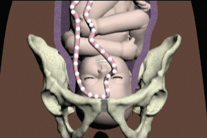
Movement of the fetus with the shifting of its position occurs to some extent in every labor. Shifting of position combined with fetal movement through the birth canal can result in umbilical cord prolapse. In this example, the cord has prolapsed over the fetal head. This type of prolapse is known as occult because the umbilical cord can not be detected on vaginal examination. Because early detection on vaginal exam is not possible with an occult prolapse, its presence may only be detected once critical impairment of blood flow to the fetus has occurred.
Just to reinforce this concept, here is an image of an overt umbilical cord prolapse, where the cord is present in the vagina and can be detected on examination. Once prolapse has occurred, uterine contractions may now compress the umbilical cord against the fetal head. Compression of the cord reduces blood flow to the fetal heart and brain. This results in a reduction in fetal heart rate, also known as a deceleration. These decelerations can be seen on the FHR monitoring strip as shown here. The decelerations can also be heard via fetal Doppler.
When the uterus relaxes between contractions, umbilical cord compression is relieved and blood flow to the fetus is restored. With the return of normal blood flow to the fetus, the deceleration resolves and heart rate returns to normal. Here’s what it sounds like on fetal Doppler. As long as there is no change in the relative position of the umbilical cord, the fetal head, and the pelvis, the process repeats. Eventually, recurrent impairment of blood and oxygen flow to the fetus may begin to have a greater physiological effect with deeper and more prolonged decelerations occurring with uterine contractions as shown here.
As the fetus advances through the birth canal, the umbilical cord becomes trapped between the head and the bony pelvis. Compression of the umbilical cord against the pelvis results in a severe reduction in blood flow to the fetus, also known as ischemia. Compression of the umbilical cord and fetal ischemia results in a sudden, severe, and prolonged deceleration of the heart rate. This makes for a dramatic change in the fetal Doppler audio. Ischemia, a severe impairment of blood flow to the fetus means that the delivery of oxygen via that blood flow is also severely impaired, a condition known as hypoxia. Ischemia and the hypoxia that results from it are the root causes of fetal hypoxic-ischemic encephalopathy.
When there is severe impairment of blood and oxygen flow due to umbilical cord prolapse, all fetal organs are affected. To maintain normal functions the brain tends to be more dependent on a steady flow of richly oxygenated blood than other organs. As a result, the brain is more sensitive to the damaging effects of hypoxia-ischemia than other organs, and when an injury occurs, it is more likely to be irreversible. The general term used to describe disease-based injury to the brain is encephalopathy.
In acute hypoxic, ischemic encephalopathy with umbilical cord prolapse, and compression, any portion of the brain or the entire brain may be injured. Areas of injury commonly seen on MRI include the thalamus, the basal ganglia, the posterior limbs of the internal capsule, and the watershed areas of the left and right cerebrum.
Cal Shipley, M.D. copyright 2021

