Lumbar Discectomy and Fusion Transcript
Lumbar Discectomy and Fusion
This is Dr. Cal Shipley with the review of discectomy and fusion in the lumbar spine.
Anatomy
Taking a look first at the anatomy, we see the fourth and fifth lumbar vertebra, or L4 and L5 as they’re known for short, and the first sacral vertebra or S1, and then the L4 L5 discs sitting between the fourth and fifth lumbar vertebra, and the L5-S1 disc sitting between the fifth lumbar and first sacral vertebra. The intervertebral discs are made of a firm but flexible material that act as shock absorbers between the spinal vertebra.
Situated as they are at the bottom of the spine, the L4, L5, and S1 vertebra and their accompanying discs bear considerable stress as a result of the weight and movement of the upper body, and as a result, have a greater frequency of injury and degenerative change on their counterparts higher up in the spine. The exception to this rule being the vertebra of the neck.
Finally, we see the spinal nerve roots exiting to the left and right between the vertebra.
Spinal Cord and the Cauda Equina
Rotating to a view from above of L5, the anatomical relationships of all these structures become a little more readily apparent. Occupying the spinal canal at this level of the spine is the cauda equina. The spinal cord ends at approximately the level of the second lumbar vertebra. The cauda equina originates from the end of the spinal cord, distributing nerve roots to the remaining lumbar and sacral vertebral levels. The left and right spinal nerve roots branch off of the cauda equina. The intervertebral disc sits on top of the vertebra and therefore between the L5 and L4 vertebrae.
Disc Degeneration
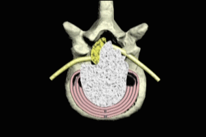
Degeneration of the disc with age or injury due to trauma may cause the disc to bulge. Depending on the severity and direction of the bulge, the spinal nerve roots or the nerves of the cauda equina may be compressed, as in this example, resulting in numbness, pain or weakness in the tissues and muscles supplied by the compressed nerves. In affected individuals, removal of the disc or discectomy, can resolve the symptoms eliminating pain and restoring normal sensation and muscular or organ function to the affected areas.
Lumbar Discectomy and Fusion Surgery
There are a variety of approaches and tools that can be utilized for disc removal. Which approach and which tools are used depends largely on the degree and direction of the disc bulge rupture and which nerve structures are being compromised, and whether or not there is abnormal growth or swelling in the bone structure of the spinal canal itself, or in the bones of the grooves through which the spinal nerves run.
Lumbar Discectomy and Fusion Anterior Approach
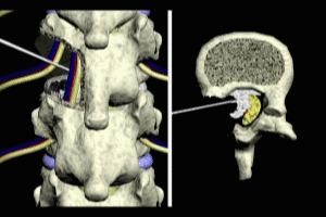
In this case, an anterior approach is being used, that is, the spine is being approached from the front of the patient. In order to perform the anterior approach to the spine, an incision is first made in the abdomen, and the various muscles, organs, and vascular structures are temporarily moved out of the way in order to provide access.
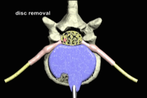
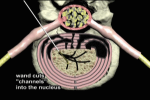
There are a variety of different tools that may be used to remove the disc. Everything from manually operated grasping and biting tools to electric drills. The choice of which specific tool or tools to use in a given case is dependent on the approach, operator preference, and of course, the specific abnormality encountered in the disc and bony structures. In this example, a grasping and biting tool known as a rongeur is used to remove the disc. The operator uses the rongeur to remove small pieces of disc, working from front to back, until the disc has been completely removed to achieve, in this case, decompression of the cauda equina.
Vertebral Gaps after Discectomy
Removal of the disc relieves pressure on the nerves or cauda equina, however, it also leaves a gap where the disc used to be between the two vertebrae. In this particular case, discs were removed at both the L4,5 and L5 and S1 levels, and so there are two gaps between the three vertebrae, as shown here. After discectomy, these gaps between the vertebra may be as much as a quarter of an inch and were they not to be filled with an alternative material, would result to an extreme instability of the spine.
Intervertebral Implants and Fusion
Currently, the most commonly used solution for filling these gaps is to place an implant device. Intervertebral implants are commonly composed of a material known as PEEK, short for poly-ether-ether ketone. Implants composed of PEEK are strong, lightweight, and do not stimulate an abnormal immune response in the recipient. Intervertebral implants contain hollowed out spaces into which bone graft material is placed to stimulate fusion between the two vertebrae. Most commonly, the graft material is termed an allograft, that is bone which has been removed from cadavers or taken from live patients during the course of joint replacement surgeries. Alternatively, the bone graft material may be autologous, that is, from the patient undergoing the spinal surgery, or composed of a synthetic material.
Implant fixation
After insertion into the intervertebral space, the implant is fixated with special screws which anchor it in place. The bone graft material stimulates additional growth of bone between the vertebrae. Six to 12 months after surgery, the fusion is complete. What were three individual vertebrae have now fused into one solid bony structure.
The fusion restores mechanical stability to the spine and greatly reduces or eliminates the pain associated with movement of vertebrae which have been subject to degenerative change.
Cal Shipley M.D., copyright 2020


