Gastrointestinal Physiology in the Extremely Premature Infant PED031
Gastrointestinal Physiology in the Extremely Premature Infant Transcript
Gastrointestinal Physiology in the Extremely Premature Infant
This is Dr. Cal Shipley with a review of gastrointestinal physiology in the extremely premature infant.
Abdominal Anatomy
Let’s start by looking at some basic anatomy. The liver is located in the right upper portion of the abdomen with the small intestine below. The diaphragm, which enables expansion and contraction of the lungs, sits between the abdomen, and the heart and lungs. Under normal conditions, the upper abdominal organs are able to accommodate the movement of the diaphragm. However, in disease states where there may be increased pressure within the abdominal cavity, the work of the diaphragm, and therefore the work of breathing, becomes much more difficult.
The colon connects the small intestine to the rectum and anus.
The Peritoneum
The liver, intestines, and most of the abdominal organs are enclosed by a membrane known as the peritoneal membrane or peritoneum. Looking at a right cross-sectional view of the abdomen, the peritoneum appears in dark blue. As can be seen, the peritoneum encloses the abdominal organs, providing a continuous membranous attachment which helps to stabilize the organs within the abdomen and prevent twisting, especially at the colon and small intestine. Within its folds, the peritoneum also carries most of the blood supply to the abdominal organs.
The peritoneum also plays a critical role in the movement of fluids and solutes within the abdominal cavity.
The Small Intestine: Peristalsis
The small intestine generates waves of muscular contractions to propel food through its length. This process is known as peristalsis. In the extremely premature infant, peristalsis may be significantly impaired, with decreased ability to propel food. The small intestine in such infants may undergo periods of ileus during which peristalsis is completely absent. In the healthy term infant, the gut is much more active with normal peristalsis.
The Small Intestine: Retention of Feeds
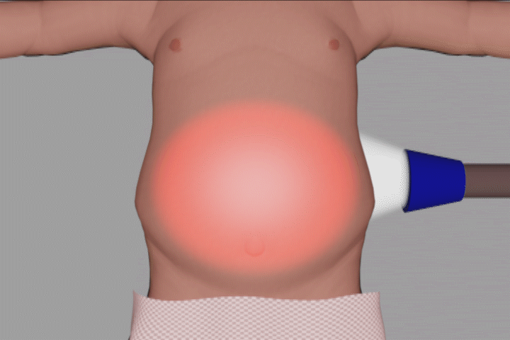
As a result of the impaired peristalsis in very premature infants, the movement of oral nutrition through the gut is slowed, a phenomenon known as retention of feeds. Extremely premature infant feeds typically take two to five days to transit through the gut. This is in sharp contrast to healthy term infants where transit time is 4 to 12 hours. Retention of feeds may result in periodic abdominal bloating or distension.
The Small Intestine: Microscopic Structure
Let’s focus now on the physiology of the small intestine at the microscopic level in a very premature infant.
Villi
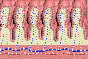
The innermost wall of the small intestine is covered by a regular layer of densely packed polyploid structures known as villi. In this depiction, the row of villi closest to us is seen in cross-section as are the other layers of the intestinal wall.
Moving in for a closer look reveals the arteries which supply oxygen and nutrients to the villi. In the extremely premature infant, the arteries are underdeveloped, which results in a reduced rate of oxygen and nutrient flow to the villi. By contrast, oxygen and nutrient flow to the villi is much more vigorous in the healthy term infant.
Epithelial Cells
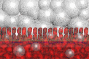
Moving in closer reveals the epithelial cells, which line the outermost aspect of each villus. The epithelial cells represent a critical physical barrier in preventing bacteria and toxins from entering the intestinal wall. Extremely premature infants have epithelial cell junctions which are looser, making it easier for bacteria and toxins to enter the intestinal wall. By contrast, healthy term infants have tight epithelial cell junctions which are much more resistant to the passage of bacteria and toxins into the intestinal wall.
Mucin
The villi of the small intestine are coated with a layer of mucin, a glycoprotein which is 95% water.
Mucin provides an additional physical barrier to harmful bacteria and toxins. In addition, it produces compounds which are active against harmful bacteria. Mucin also prevents autodigestion of the intestinal wall by digestive enzymes. In the extremely premature infant, the mucin layer is underdeveloped and much thinner than that of a healthy term baby.
Commensal Bacteria
In the healthy term infant, helpful bacteria, known as commensals, interact with epithelial cells to develop immune defenses, produce mucin, and improve digestion and peristalsis. In the extremely premature infant, the population of helpful bacteria is underdeveloped and sparse, and antibiotics administered to treat the growth of harmful bacteria may further reduce the population of commensals.
Cal Shipley, M.D. copyright 2020


