Necrotizing Enterocolitis (NEC) Transcript
Necrotizing Enterocolitis (NEC)
This is Dr. Cal Shipley with a review of necrotizing enterocolitis, as may occur in an extremely premature infant of 24 weeks gestation.
Abnormal Bacterial Colonization in Necrotizing Enterocolitis
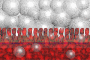
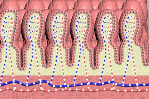
Let’s start our review of necrotizing enterocolitis by looking at a cross-sectional view of the small intestine, focusing on the intestinal wall. We’ll zoom in for a view at the microscopic level. The inner wall of the small intestine, also known as the mucosa consists of millions of polyploid structures known as villi seen here in cross-section. Because virtually 100% of premature 24-week gestation infants remain in hospital after birth, their intestinal tracts are colonized by hospital-based bacteria, many of them harmful pathogens.
In addition, bacteria from the infant’s colon may move up into the small intestine. Under normal circumstances, these colonic bacteria may not be pathogenic, however, their presence in the small intestine in large numbers may cause problems such as excessive gas formation.
Impaired Intestinal Peristalsis
Very premature infants have an underdeveloped ability to move food through their intestinal systems, resulting in retention of feeds. The prolonged presence of food in the gut provides a rich substrate for the growth of harmful bacteria.
Underdeveloped Immunity
The underdeveloped defenses associated with extreme prematurity are unable to control rapid reproduction of the harmful bacteria.
Inflammation and Gas Formation in Necrotizing Enterocolitis
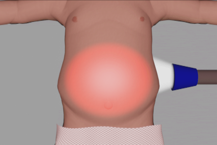
As the bacterial population increases, the wall of the intestine becomes inflamed. Overgrowth of gas-forming bacteria results in production of gas bubbles. Gas formation distends the small intestine and the colon. This massive gas-related distension of the intestines is known as pneumatosis intestinalis. Here is a good example on plain film X-ray of pneumatosis intestinalis in a premature infant with necrotizing enterocolitis. The gas distended intestines are outlined in blue. The intestinal distension exerts pressure on the diaphragm, impairing lung expansion, and therefore inhibiting respiration.
The gas in the intestine is under such pressure that it is forced into the intestinal wall, and into the blood vessels within the wall.
Portal Venous Gas
This cutaway view demonstrates a portion of the web of veins which drain blood from the intestine into the liver via the portal vein. Gas which has entered veins within the intestinal wall travels to the portal vein. Accumulations of gas within the portal vein may be detected on imaging studies.
Ultrasound
Ultrasound is particularly sensitive in detecting portal venous gas and may allow for early diagnosis of necrotizing enterocolitis.
Plain Film Xray
Portal venous gas may also be seen on plain film X-ray of the abdomen, however, plain film imaging is much less sensitive than ultrasound. As a result by the time portal venous gas is detected, the disease process is at a more advanced stage.
Here is a plain film X-ray of an infant with necrotizing enterocolitis, in which the portal venous gas may be seen as a string of darkened bubbles just to the left of the arrow.
Intestinal Ischemia in Necrotizing Enterocolitis
As the process continues, intestinal distension, gas, and inflammation combine to compress arteries in the intestinal wall, causing a critical reduction in blood flow known as ischemia. The ischemia causes necrosis in the intestinal wall resulting in perforations. Gas and bacteria-laden discharge leak from the perforations. Much of the leakage occurs into the peritoneum.
Peritoneal Gas and Fluid
This view of the abdomen from the right side in cross-section demonstrates the presence of gas and bacteria-laden fluid within the peritoneum. Some of the perforations will erode through the peritoneum releasing fluid and free air into the abdomen.
Septicemia
Eventually, bacteria penetrate the intestinal wall and enter the bloodstream. The presence of bacteria in the bloodstream is known as septicemia. Once in the bloodstream, the bacteria are distributed to all organs. Ultimately septicemia results in acidosis with multi-organ failure and cardiac arrest.
Cal Shipley, M.D, copyright 2020


