Chest Compression Physiology RES005
Chest Compression Physiology Transcript
Chest Compression Physiology
This is Dr. Cal Shipley with a review of the physiology of chest compressions during cardiopulmonary resuscitation.
Chest Compressions First
After confirming that the victim has no pulse, chest compressions are started. Chest compression should be started before any attempt to administer rescue breathing (formerly known as artificial respiration) either by mouth-to-mouth or Ambu-bag as shown here.
The reasoning behind this recommendation is that most individuals will have a residual amount of oxygen in their bloodstream after a cardiorespiratory arrest. The first priority in resuscitation is to circulate this oxygen to the heart and brain, the two organs whose response to a critical loss of oxygen flow has the most devastating impact on human viability. Chest compressions are the priority in resuscitation and current American Heart Association guidelines recommend 30 chest compressions be performed prior to the initiation of rescue breathing.
Chest Compression Technique
Now let’s take a closer look at exactly how chest compressions work. Let’s switch to a view of the chest from the left side. In adults, compressions of the chest are achieved by applying the heel of one hand over the lower half of the sternum, with the second hand applied to the top of the first, and with elbows locked, applying downward pressure. The chest should be compressed to a depth of at least two inches, and at least 100 compressions should be performed per minute.
Chest Compression Phases
When discussing the physiology of chest compressions, scientists recognize two important phases: compression and decompression.
Compression Phase
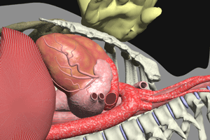
Let’s start with the compression phase. To make this easier to visualize, I’m going to peel back the layers of the chest. Here are the left ribs, the left lung, and the pericardial sac, which anchors the heart within the chest; the sternum, or breastbone, and as you’ll note, the hand is applied over the lower half as previously mentioned.
Removal of the pericardium reveals the heart. From this viewpoint, the left ventricle and left atrium are visible. The aorta originates from the left ventricle and runs behind the heart between the heart and the spine. The aorta is responsible for carrying oxygen-rich blood to all bodily organs and tissues, including the brain and the coronary arteries of the heart.
In order to be effective, during the compression phase the sternum must be depressed at least two inches, and at a rate of at least 100 compressions per minute.
Switching to a cross-sectional view of the heart reveals the blood contained within the cardiac chambers. During the compression phase, depression of the sternum compresses the heart, increasing the pressure within the cardiac chambers, which propels blood forward into the aorta.
The key to the forward movement of the blood through the heart during compression are the cardiac valves, which allow one-way flow only.
During the compression phase, blood is pushed into the aorta and up into the brain via the carotid arteries. From the aorta blood also flows into the coronary arteries.
Decompression Phase
The second phase in chest compressions is known as the decompression phase. The decompression phase of chest compressions is considered by resuscitation researchers to be of equal importance to the compression phase.
Decompression creates a negative pressure within the thorax, in essence, generating a vacuum effect within the chest cavity. This negative pressure causes the superior and inferior vena cava to expand and fill with blood, which then enters the right atrium to which they are connected. There is also a negative pressure generated within the cardiac chambers as they expand during decompression. This negative pressure draws blood into the chambers.
The decompression phase is important because it facilitates the refilling of the cardiac chambers with blood and readies the heart for the next compression.
It is critically important that the person performing chest compressions allow the sternum to completely recoil to its pre-compression position before initiating the next compression. This allows for maximum refilling of the cardiac chambers and therefore the maximum flow of blood to the brain and heart with the next compression.
Additional Considerations
I’d like to touch on just a couple of additional points before I close this presentation.
Technique
Resuscitation researchers have found that the compression rate should be at least 100 compressions per minute. They have also determined that compression rates of greater than 120 per minute do not allow a sufficient amount of time during the decompression phase for the cardiac chambers to adequately refill, thus reducing the volume of blood flow to the heart and brain.
Chest Compression in Barrel Chested Individuals
Finally, chest compressions have been found to be effective even in barrel-chested individuals who have a significant gap between the back of the sternum and the front of the heart. In these individuals, the degree of cardiac compression with chest compressions is markedly reduced. Based on the decreased cardiac compression in these individuals, one would expect blood flow as a result of CPR to be much reduced. Surprisingly, researchers have found that flow in barrel-chested individuals is in many cases comparable to that of individuals with normal thoracic dimensions.
The Thoracic Pump
This apparently contradictory finding would seem to lend support to an alternative physiological mechanism for blood flow in barrel-chested individuals undergoing chest compressions. Sure enough, scientists have recently come up with a fascinating theory called the thoracic pump mechanism. I’m not going to go into the details of this mechanism in this presentation. However, I have included a couple of links on this webpage where you can get much more information. If you’re interested in CPR physiology, these articles are definitely worth a look.
Cal Shipley, M.D. copyright 2020

