Aortic Dissection and Cardiac Tamponade CVS062D
Aortic Dissection and Cardiac Tamponade Transcript
Aortic Dissection and Cardiac Tamponade
Aortic and Cardiac Anatomy
This is Dr. Cal Shipley with a review of aortic dissection leading to acute cardiac tamponade. Let’s start by taking a look at the relevant cardiac and aortic anatomy.
The heart is positioned in the chest between the left and right lung and is enclosed by the pericardial sac. In addition to the heart, the pericardial sac encloses the distal portions of the great vessels, the superior and inferior vena cava and the aorta. The thoracic spine lies directly behind or posterior to the aorta.
The Pericardial Sac
The pericardial sac completely encloses the heart, creating a liquid-tight seal at the points of entry of the vena cava and aorta, as well as the other major cardiac vessels.
A cross-sectional view of the pericardial sac reveals the heart with the left and right coronary arteries. At the top of the aorta is a curved section known as the Arch. This portion of the aorta carries the greatest blood flow and is subjected to the highest blood pressure of any vessel in the human body.
Taking a closer look at the aortic wall reveals a three-layer structure consisting of an outer layer, the adventitia, a middle layer, the media, and an inner layer, the intima.
Aortic Dissection
Now, let’s take a look at the process of aortic dissection resulting in acute cardiac tamponade. Aortic dissection typically occurs in older adults in the presence of disease of the aortic wall, also known as atherosclerosis or hardening of the arteries.
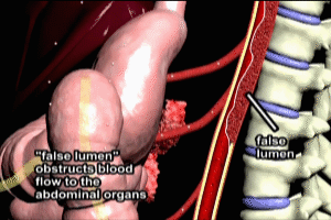
Subjected to the constant stress of the high blood pressure within the aorta, the diseased inner wall, (the intima) will develop a tear. The tear exposes the media layer. Blood flows into the tear resulting in dissection of the intima layer away from the media layer. The dissection may occur either up or down the arch, or both, creating a channel within the aorta known as a false lumen.
Cardiac Tamponade
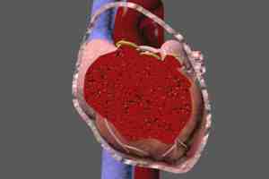
Eventually, the dissection may progress to the portion of the aorta sealed within the pericardial sac. The pressure within the false lumen may cause a rupture of the adventitia, resulting in hemorrhage of blood into the pericardial sac. Blood flows rapidly into the pericardial sac. The pericardial sac has a limited capacity to accommodate the blood volume, and when this capacity is reached, increasing pressure is exerted on the cardiac chambers. Cardiac output of blood is impaired, and to compensate, the heartbeat increases. Increasing tamponade of the chambers of the heart eventually results in cardiac arrest.
copyright 2023 Cal Shipley, M.D.


