Fetal Heart Monitoring Review OBS195
Fetal Heart Rate Monitoring Transcript
Fetal Heart Rate (FHR) Monitoring and Interpretation
This is Dr. Cal Shipley with a review of fetal heart rate monitoring during labor. Fetal heart rate monitoring is also known by the acronym FHR. FHR is the most common means of assessing the health of the fetus, also known as fetal viability during the process of labor. To better understand how FHR works, let’s take a look at some fetal and maternal anatomy.
Fetal and Maternal Anatomy
Depicted here is a pregnant or gravid uterus at term, that is, carrying a fetus with a gestational age of approximately 40 weeks. Switching to a cross-sectional view of the uterus, we see the fetus contained within, and attached to the wall of the uterus, the placenta. Connecting the placenta to the fetus is the umbilical cord containing the umbilical arteries and the umbilical vein.
Rotating the uterus 180 degrees, and switching to an internal view of the fetus, reveals the fetal heart, with the umbilical cord connecting the placenta to the fetal circulatory system. The maternal uterine artery sends branches through the uterine wall into the placenta. Oxygen and nutrient rich blood flows from the uterine artery through the placenta and umbilical cord to all fetal organs and tissues. This of course includes the brain, which regulates the fetal cardiac activity. Thus, the fetus is completely dependent upon the mother for nutrition, and cannot survive without a steady flow of blood through the umbilical cord.
Fetal Hypoxemia
Significant impairments or interruptions in blood flow to the fetus will, therefore, result in lower than normal blood levels of oxygen, also known as hypoxemia. The usefulness of fetal heart rate monitoring during labor is based on the fact that significant or prolonged hypoxemia will result in FHR pattern changes, particularly decreased heart rate, also known as bradycardia, and loss of fetal heart rate variability. I’ll be discussing the meaning of loss of variability a little further on in the presentation.
Significant changes in FHR patterns may also occur with loss of fetal blood volume, also known as hypovolemia or fetal acidemia. In both of these instances however, FHR changes are related to hypoxemia. In the case of hypovolemia, decreased circulating blood volume leads to decreased levels of oxygen in the blood stream and in acidemia, the acid based disturbances are most commonly a result of prolonged hypoxemia.
Fetal Heart Rate Monitoring Techniques, Normal Heart Rate, Variability
Now, let’s turn to how monitoring of the fetal heart rate is actually accomplished. The most common method used to record the fetal heart rate, as well as uterine contractions, is to apply two external sensors to the mother’s abdomen. The heart rate sensor is applied in the area of the fetal heart, with the uterine contraction sensor typically placed above it. The wall of the gravid uterus contains a thick muscular layer capable of generating significant force as it propels the fetus through the birth canal.
Normal Fetal Heart Rate
During labor, normal uterine contractions appear as wave-like forms, as seen here on this actual FHR recording strip. In contrast, a normal fetal heart rate tracing looks more like a squiggly line. During labor, a normal fetal heart rate is approximately a 110 to a 170 beats per minute.
Fetal Heart Rate Variability
In a normal fetal heart rate recording, the squiggly characteristic of the tracing results from random variations in the heart rate also known as variability. Normal fetal heart rate variability is about 6 to 25 beats per minute. Variability within this range indicates a healthy nervous system and normal cardiac responsiveness. Loss of normal variability can be an ominous sign with respect to the condition of the fetus, and we’ll be looking at some examples of this later in the presentation. Most modern fetal heart rate monitoring equipment also outputs a sound component, changes in which can alert the delivery room staff to problems with the fetal status.
[fetal heart beat]
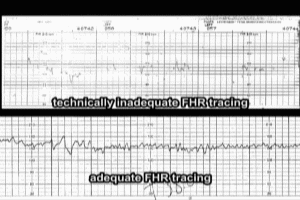
The external monitor system I have discussed thus far is generally very reliable for obtaining an adequate FHR tracing, and it has the advantage of being completely non-invasive, that is, in no way detrimental to the fetus and the mother. In some instances however, for example when there is excessive fetal or maternal movement, the external system may not yield a readily interpretable tracing.
Here is an example of a technically inadequate tracing produced by an external monitor system. Compare it to a normal tracing. The inadequate tracing would be virtually impossible to interpret. In such instances, an electrode is attached to the fetal scalp, ensuring a good quality tracing. The electrode is inserted through the vagina and cervix, and by means of a coil attached to the scalp.
Unlike external FHR monitoring, scalp electrodes are considered an invasive technique, as the tip of the coil must pierce the fetal scalp in order to be attached. Serious complications associated with fetal scalp electrodes are rare. Not uncommonly, scalp electrodes may cause bleeding within the scalp, which can cause the formation of a pocket of blood known as a hematoma. While scalp hematomas may be unsightly, they generally resolve spontaneously within several days of birth, and cause no further complications.
Fetal Heart Rate Decelerations
Now that we know a little bit more about what a normal FHR tracing looks like, and how it is obtained, let’s take a look at some variations, specifically decelerations, loss of variability and bradycardia. Let’s start by looking at the normal FHR tracing, with the fetal heart rate within the normal range of a 110 to a 170 beats per minute, and heart rate variability within the normal range of 6 to 25 beats per minute.
As a side note, let me mention that these values for heart rate and heart rate variability are not universally agreed upon, and may vary slightly depending on the source. Again, as mentioned previously, along the lower half of the FHR strip is the uterine contraction tracing. This particular tracing is consistent with active labor, with normal intensity contractions occurring approximately every two minutes.
The term deceleration refers to a decrease in the fetal heart rate of at least 15 beats per minute from baseline. In addition, the timing of the decelerations relative to uterine contractions is important in their interpretation.
[fetal heartbeat]
Let’s start with our review of decelerations by looking at early decelerations. Early decelerations have a slow onset and a uniform shape and their nadir, the point at which the fetal heart rate is at its lowest point, corresponds with the peak of uterine contractions as seen here. The fetal heart rate returns to baseline with the end of the uterine contraction, and normal beat-to-beat variability is maintained throughout.
Early decelerations are caused by compression of the fetal head during uterine contractions. This compression causes stimulation of the vagus nerve, which temporarily reduces fetal heart rate. Early decelerations are not considered to be a sign of fetal distress, and as such, are consistent with a reassuring fetal heart rate pattern.
[fetal heartbeat]
Now let’s consider late decelerations. Late decelerations consist of a symmetrical pattern of heart rate deceleration, which begin at, or after, the peak of uterine contraction. Late decelerations are associated with uteroplacental insufficiency, and are triggered by uterine contractions. In essence, this means that any condition which impairs uterine blood flow, or results in a significant decrease in placental function, can cause late decelerations.
Conditions which can give rise to decreases in uterine blood flow include, abnormally low maternal blood pressure, also known as hypotension, uterine hyperstimulation, most commonly seen with induction of labor, and the acidosis, and decreased blood volume (also known as hypovolemia), which may occur in mothers with poorly controlled diabetes mellitus. Causes of decreased placental function include post-date gestation, preeclampsia, chronic maternal hypertension and maternal diabetes.
Persistent late decelerations are always considered to be non-reassuring, and should trigger evaluation of fetal pH to look for acidosis. Here’s an example of deep late decelerations with loss of variability. Note how smooth the tracing line is compared to normal. The nadir of each deceleration reaches 90 to 100 beats per minute, well below the normal level. Loss of variability, and the presence of persistent late decelerations, is an ominous sign, and should prompt serious consideration for immediate delivery.
[fetal heartbeat]
The last category of decelerations we are going to look at are variable decelerations. Variable decelerations are the most commonly encountered decelerations during labor. They are caused by intermittent compression of the umbilical cord during uterine contractions, and are particularly seen with mothers who have had premature rupture of membranes and decreased amniotic fluid volume.
During cord compression, occlusion of the umbilical artery causes a sharp downslope in the fetal heartbeat, with a prompt return to baseline, and often a brief acceleration, indicating a healthy response. This results in a pattern which is often characterized as V or U shaped.
The onset of variable decelerations may vary with respect to the timing of uterine contractions, hence their name. They may be classified according to their depth and duration as mild, moderate or severe. Here is an example of a moderately severe variable deceleration pattern with depth 70 to 80 beats per minute, and duration approximately 25 to 30 seconds. A persistent variable deceleration pattern in the fetal heart tracing is a non-reassuring sign and may result in fetal acidosis and distress.
This particular tracing also exhibits loss of variability. As with persistent late decelerations, loss of variability in variable deceleration patterns is an ominous sign, and should prompt immediate corrective measures or delivery. Putting all this together, let’s take a look at how variable decelerations occur with respect to umbilical cord compression and uterine contraction in real time. Prior to contraction, blood flows freely through the umbilical cord from the placenta to the fetus.
[fetal heartbeat]
As contraction proceeds, the umbilical cord is compressed between the fetal head and the uterine wall impairing blood flow.
[fetal heartbeat]
As the urine contraction ends, blood flow through the umbilical cord is restored.
[fetal heartbeat]
Fetal Heart Rate Bradycardia
The term bradycardia refers to decreased heart rate, and it is defined by the American College of Obstetrics and Gynecology as a baseline fetal heart rate of less than a 110 beats per minute. Moderate bradycardia of 80 to 110 beats per minute is a non-reassuring sign. Loss of variability during moderate bradycardia, or severe bradycardia, defined as heartbeat of less than 80 beats per minute, occurring for three minutes or longer, are ominous signs, and indicates severe fetal hypoxia. Causes of severe fetal bradycardia during labor include umbilical cord compression, particularly in association with umbilical cord prolapse.
Here’s an example of umbilical cord compression occurring during labor as a result of a low lying or occult prolapse of the umbilical cord. The term occult refers to the fact that the umbilical cord is contained completely within the uterus, and cannot be seen or felt by the examining physician. The cord becomes trapped between the fetal head and the wall of the uterus, and with the progression of labor, may become completely compressed, cutting off all oxygen flow to the fetus.
Other causes of severe fetal bradycardia during labor include, titanic uterine contractions usually in association with induction of labor, paracervical anaesthetic block, epidural and spinal anesthesia, maternal seizures, rapid descent of fetus through the birth canal, and vigorous vaginal examination.
[fetal heartbeat]
Fetal Heart Rate Accelerations
Finally, let’s take a look at accelerations of the fetal heart rate and fetal tachycardia during labor. An acceleration is defined as an increase in the fetal heart rate, where the increase from onset to the peak of the acceleration is less than 30 seconds, greater than 15 beats per minute above the baseline, and with a duration of at least 15 seconds from onset to return. Fetal heart rate accelerations are almost always benign and reassuring. They may occur periodically, that is, in relation to the uterine contractions, and usually occur due to fetal stimulation, especially with breech presentation or when the umbilical cord is compressed with compression of the umbilical vein only.
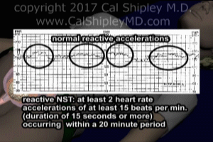
Episodic fetal heart rate accelerations occur independently of uterine contractions and are usually due to fetal movement. Episodic accelerations are also a normal response to fetal scalp stimulation. Episodic accelerations are also one of the three criteria that form the basis of the non-stress test. A non-stress test is said to be reactive, or normal, when two episodic accelerations occur over a 20 minute period, with moderate variability, and no decelerations in the fetal heart rate tracing.
The FHR chart tracing depicted here would be considered reactive. Fetal tachycardia is said to occur when the baseline heart rate exceeds a 160 beats per minute, as depicted here. Fetal tachycardia during labor is considered to be a non-reassuring sign and has multiple potential causes as listed here.
Cal Shipley, M.D. copyright 2020


