Anterior Lumbar Interbody Fusion (ALIF Part 3) ORT073
Anterior Lumbar Interbody Fusion Transcript
Anterior Lumbar Interbody Fusion
This is Dr. Cal Shipley with anterior lumbar interbody fusion part three: ALIF and posterior laminectomy and hardware placement.
Spinal Disease Model
Here is a view of the diseased lumbar spine that we are going to be using in this presentation. The intervertebral discs at L3-4 and L4-5 have completely collapsed, while the L5-S1 disc is partially collapsed.
Previous Spinal Surgery
Let’s rotate our view 180° so we can see the back of the spine. It’s fairly common for patients with multi-level disc disease to have had spinal procedures performed at earlier stages of their disease.
The vertebral laminae are areas of bone which sit on either side of the spine as depicted here.
In this example, the patient has had a previous right-sided hemilaminectomy with removal of the L3, L4 and L5 laminae on the right side of the spine only. The hemilaminectomy creates an opening into the spinal canal and exposes the cauda equina (I’m going to discuss the purpose of a laminectomy later in the presentation.)
Anterior versus Posterior Spinal Approach
The previously performed hemilaminectomy causes adhesions to form between the cauda equina and the disc spaces. The scarring makes it both difficult and dangerous to use a posterior approach when performing spinal surgery to treat multi-level degenerative disc disease. The surgeon’s ability to see the cauda equina clearly and keep it out of harm’s way is significantly impaired, and the risk of neurological injury too great. In these circumstances, the anterior spinal approach is much preferred.
Anterior Lumbar Interbody Fusion Procedure
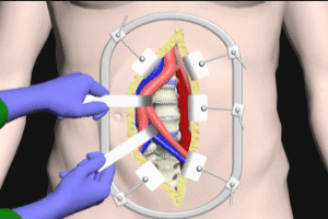
Once the spine has been exposed by the retroperitoneal approach, as detailed in part two of this series, the surgeon may proceed with the interbody fusion.
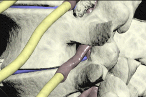
The L5-S1 disc is removed and the disc space is prepared for the interbody device. The interbody device, also known as a fusion cage, is typically made of carbon fiber or titanium and has an open center to allow for the placement of graft material. Grooving helps to keep the device in place after insertion.
Graft Material
Typically, the graft material is autograft, bone that is harvested from elsewhere in the patient’s body, or allograft, graft material donated from another individual. For more in depth review of bone graft materials and recently developed alternatives, please see the helpful links on this page.
Interbody Device Placement
The interbody device is now inserted into the disc space and anchored in place with screws, which enter the bodies of the L5 vertebra above and the S1 vertebra below.
Moving up to the L4-5 disc space level, accessible bone growths and spurs are removed and then a special tool is used to open up the collapsed disc space. The disc remnant is removed, the disc space is prepared, and then an interbody device packed with graft is inserted into the disc space and anchored with screws.
The same technique is used to place an interbody device at the L3-4 disc space. The ALIF portion of the procedure is complete.
Results
The placement of the interbody devices restores the openings of the neural foraminae and relieves nerve root compression.
The lumbar spine bears the load of the upper body weight, compressing the vertebrae against the interbody devices and providing stability.
Posterior Laminectomy Procedure
Let’s take a look now at a posterior laminectomy, with placement of hardware. This is a procedure which often accompanies an anterior lumbar interbody fusion.
Spinal Disease Treatable from a Posterior Approach
As mentioned earlier in the presentation, individuals affected by degenerative disc disease at multiple levels often have significant degenerative bone spurs and bone growth along the posterior aspect of the vertebrae and disc spaces, causing narrowing of the spinal canal and compression of the cauda equina.
It may not be possible to adequately treat this bony material using an anterior approach, and so immediately after the ALIF procedure has been completed, a posterior approach is used to perform laminectomy.
Posterior Laminectomy
Let’s turn the spine 90° so that we can view it from behind. As mentioned previously in the sample case that I’m using today, the patient had had a previous right-sided hemilaminectomy at the L3, 4 and 5 levels to deal with the degenerative disc disease at an earlier stage. Removal of the left-sided lamina, as well as the spinal bone, converts the situation into a full laminectomy. Viewing the spine from the left side in cross-section reveals the decompression of the cauda equina as a result of the full laminectomy.
Placement of Stabilization Hardware Post-laminectomy
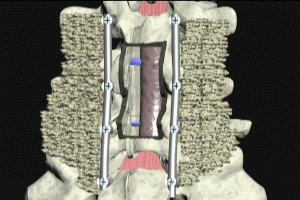
One consequence of the multi-level full laminectomy is potentially a destabilization of the spine at the L3, 4 and 5 levels. To remedy the situation, supporting hardware is attached to the spine. Screws are inserted into the pedicles of the vertebrae. A metal rod, known as a strut, is then affixed to the pedicle screws by threaded caps. This same procedure is followed to attach a strut to the opposite side.
Graft Fusion
The graft material placed into the disc spaces require several months to fuse into solid bone.
While the graft is fusing, the interbody devices and attached hardware provide a stable construct able to bear upper body load without shifting. Lateral flexion of the spine, as well as anterior/posterior flexion and extension are supported by the hardware, while the graft material undergoes fusion.
Once fusion is complete, the construct provides a stable and sturdy long-term platform for the spine above. The procedure also restores the normal curvature of the lumbar spine. Normal alignment of the vertebrae is also restored.
Cal Shipley, M.D. copyright 2020
