Retroperitoneal Exposure of the Lumbar Spine (ALIF Part 2) ORT072
Retroperitoneal Exposure of the Lumbar Spine Transcript
Retroperitoneal Exposure of the Lumbar Spine
This is Dr. Cal Shipley with part two in my four part series on Anterior Lumbar Interbody Fusion.
In this presentation, I’m going to look at the procedure to expose the lumbar spine using the retroperitoneal approach. Let’s start by looking at some abdominal anatomy.
Abdominal Anatomy
The Peritoneum
The abdominal organs are enclosed within the peritoneal membrane. Let’s look at a cross-sectional view of the abdomen from above. The peritoneal membrane, here shown in yellow, encloses the small intestine. The peritoneal membrane also encloses the liver, most of the stomach, and most of the large intestine.
The Retroperitoneum
Removing the small intestine reveals the back wall of the peritoneal membrane. Structures such as the spine or pancreas, which lie behind the back wall of the peritoneal membrane, are termed retroperitoneal.
The Aorta and Vena Cava
The largest vein in the body, the vena cava and the largest artery, the aorta, are both retroperitoneal structures. Looking again at a cross-sectional view of the abdomen from above, the aorta and vena cava lie behind the peritoneal membrane and against the lumbar spine. Because of their anatomical location, both the vena cava and the aorta must be moved away from the lumbar spine in order for the interbody fusion to take place.
Retroperitoneal Lumbar Spine Exposure Procedure
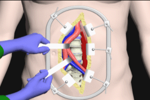
Now turning to the spinal exposure procedure – typically, a left paramedian abdominal incision is made down to the peritoneal membrane. The peritoneal membrane is left intact and is neither cut nor entered. The surgeon uses a blunt dissection with her hand to gently separate the peritoneal membrane from the inner backside of the abdominal wall. The peritoneal membrane, and the organs that it contains, are swept to the side to expose the aorta, vena cava and the lumbar spine.
Retroperitoneal Spinal Exposure Advantages
This retroperitoneal approach avoids cutting or entering the peritoneal membrane and significantly reduces the risks of a post-operative abdominal infection.
The retroperitoneal technique also avoids direct manipulation of the intestines and their blood supply reducing the chance of perforation, rupture, infection or hemorrhage.
The older transperitoneal approach requires cutting through the peritoneal membranes with extensive manipulation of the intestines and their blood supply.
The retroperitoneal approach does not cut the peritoneal membrane, minimizes intestine and blood supply contact, and reduces risks overall.
Lumbar Disk Space Access
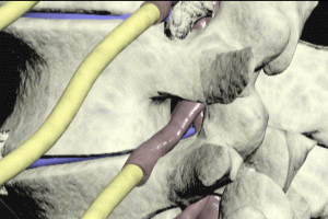
After exposure with the aorta and vena cava in their usual position, the L5-S1 disk space may be accessed by the surgeon. Unlike the L5-S1 disk level, the L3-4 and L4-5 vertebral and discs are hidden behind the aorta and vena cava. In order to access these levels, the vena cava and aorta must be repositioned by the surgeon. Handheld ribbon retractors gently pull the vena cava, aorta and the left iliac artery and vein to the right. The L3-4 and L4-5 disc spaces are now accessible, and the ALIF procedure may proceed.
Cal Shipley, M.D. copyright 2020


