Degenerative Disc Disease Lumbar (ALIF Part 1) ORT071
Degenerative Disc Disease Transcript
Degenerative Disc Disease
This is Dr. Cal Shipley with Anterior Lumbar Interbody Fusion Part 1: Spinal Anatomy and Degenerative Disc Disease.
Spinal Anatomy
Intervertebral Discs
In this case, we’re going to be focusing on the third, fourth, and fifth lumbar, and first sacral vertebrae. In between each pair of vertebrae are the intervertebral discs, which allow for movement of the vertebrae with respect to each other, and act as shock absorbers for mechanical stress on the spinal column.
Neural Foraminae and Spinal Canal
Rotating the spine 90 degrees to a side view reveals the neural foraminae openings through which pass nerve roots from the spinal cord. A cross-sectional view reveals the spinal canal, which at this level no longer contains the spinal cord, but rather, the bundles of nerve roots coming from the tail of the spinal cord, also known as the cauda equina (Latin for horse’s tail).
Disc Anatomical Relationships
Now, let’s return to the frontal view of the spine and more closely examine the L5 vertebra and its anatomical relationships. Looking from above, the L4-L5 intervertebral disc sits atop the vertebral body. The spinal canal is located directly behind the vertebral body. Within the canal is the nerve bundle, which is the cauda equina, from which originates both a left and right spinal nerve root.
Degenerative Disc Disease
Now, let’s turn our attention to degenerative disc disease. The underlying cause of disc degeneration is a reduction in the number of healthy disc cells being produced within the disc. In turn, the decreased production of healthy disc cells appears to be related to multiple factors, including aging, smoking, deficiency of nutrients & oxygen within the disc (which appears to be aggravated by both aging and smoking), genetic predisposition, and mechanical loading, particularly in the lower lumbar spine, which is a focal point for stress transmitted from the spine above.
Disc Collapse
As the process of degeneration progresses, discs collapse under the load of the spine above. In advanced cases, this results in extreme narrowing of the disc space. Severe loss of intervertebral spacing, as shown here, can result in a bone-on-bone situation in which the vertebrae are in direct contact with each other. This direct vertebral contact results in significant friction as the vertebrae move with respect to each other during changes in spinal position. Friction, in turn, can result in inflammation and pain. Loss of intervertebral spacing can also narrow neural foraminae, resulting in compression of nerve roots.
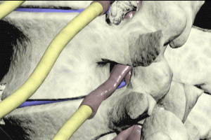
Here’s a closeup view of the entrapment and compression of a spinal nerve root due to a narrowed neuroforamen.
Vertebral Degeneration
In a majority of cases, individuals affected by degenerative disc disease have accompanying degeneration of the vertebra, resulting in abnormal bone growth and spurs, which may further compromise spinal nerve roots in the neural foraminae. Other bone growths and spurs are commonly found elsewhere on the vertebrae and may result in ankylosis, as shown here, in which two vertebrae may become fused together by a bone growth.
Disc Dessication and Spinal Canal Compression
In this example, the L3-L4 and L4-L5 discs are completely collapsed while the L5-S1 disc shows signs of partial collapse or flattening as well as desiccation, a term which describes the loss of normal water content in the disc.
Here is what the abnormalities we’ve been discussing look like on an MRI image. In addition to the loss of normal intervertebral spacing, collapsed discs commonly bulge or herniate into the spinal canal, compressing the nerve fibers of the cauda equina.
Viewing the L4 vertebra from above gives us a good angle on how spinal canal compression occurs in the early stages of disc degeneration. The disc bulges into the cauda equina, causing mild compression. As degeneration and collapse progress, the nucleus of the disc may herniate through its outer shell, resulting in moderate to severe compression of the cauda equina.
Over time, the herniated contents may calcify, and the process of calcification may expand, compressing the spinal nerve roots.
Multilevel Degeneratve Disc Disease
This same process may occur at all levels affected by the degenerative disc disease. Multilevel degenerative disc disease may also cause alterations in the normal curvature of the spine, as demonstrated here. The alignment of the spinal vertebrae may also be affected, as shown here.
Cal Shipley, M.D. copyright 2020


