 FETAL SHOULDER DYSTOCIA: DEFINITION
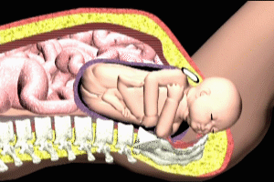
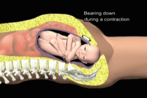
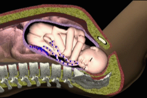
During the process of human birth, the term Shoulder Dystocia refers to the entrapment of the fetus within the birth canal. This entrapment results from the impaction of either the Anterior Shoulder (most common) against the maternal pubic bone, or the Posterior Shoulder (least common) against the bony protuberance of the sacral bone (see animation of maternal-fetal anatomy). Occasionally, both shoulders may become impacted. Prolonged or severe shoulder dystocia may lead to serious injury of the plexus of nerves supplying sensation and function to the fetal upper limb, and can also result in permanent brain damage as a result of interruption of fetal cerebral blood flow. Fetal Shoulder Dystocia may occur in up to 3% of all vaginal deliveries, and it is critical that delivery physicians and staff be well versed in both the recognition and treatment of this problem.
Most authorities have defined significant shoulder dystocia to include any delivery requiring one or more maneuvers (discussed below) in addition to gentle downward traction on the fetal head to effect delivery.
RELEVANT MATERNAL AND FETAL ANATOMY
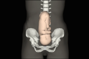
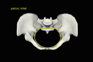
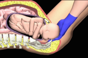
The key maternal anatomical elements in shoulder dystocia are the pubic bone, which forms the anterior (frontal) border of the pelvic inlet, and the sacrum, which forms the posterior (rear) border of the inlet.
MATERNAL FETAL ANATOMY
FETAL SHOULDER DYSTOCIA: DYSTOCIA OF THE ANTERIOR SHOULDER
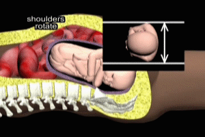
When the fetal shoulders fail to adequately rotate upon reaching the pelvic inlet, the anterior fetal shoulder may become impacted on the maternal pubic bone. This is by far the most common form of fetal shoulder dystocia. The shoulder may spontaneously dislodge with further uterine contractions, without harm to the fetus. Many such cases of shoulder dystocia likely go unnoticed by the delivery staff. If, however, the shoulder impaction persists, fetal injury may occur.

DYSTOCIA OF THE POSTERIOR SHOULDER
Dystocia of the posterior shoulder may also result from inadequate rotation of the fetal shoulders as they enter the pelvic inlet. In this case, the posterior shoulder (nearest the mother’s spine) impacts against the promontory of the sacrum. This is the less common form of dystocia.
FETAL SHOULDER DYSTOCIA: FETAL INJURIES
BRACHIAL PLEXUS INJURY
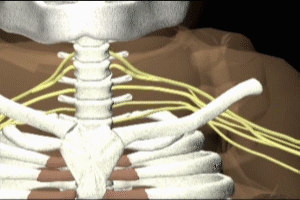
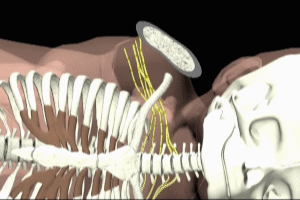
The most common injury associated with shoulder dystocia is injury to the brachial plexus. The brachial plexus is a group of nerves located in the lower neck, formed by 5 nerve roots emerging from the spinal cord as it runs through the cervical spine.
The mechanism of plexus injury resulting from operator traction on the the fetal head and neck in attempts to deliver an infant whose shoulder is impacted is controversial. Many researchers are skeptical of this mechanism, and point to the fact that 40% of all brachial plexus injuries occur in deliveries without known shoulder dystocia, where no undue force is exerted on the fetal head.
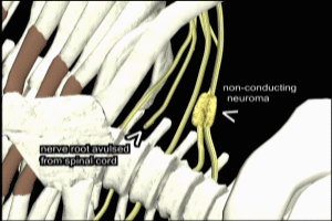
Most commonly, the nerves of the C5 and C6 nerve roots are injured, resulting in the clinical findings of Erb-Duchenne palsy: the infant loses the power to abduct the arm from the shoulder, rotate the arm externally, and supinate the forearm. The characteristic position consists of adduction and internal rotation of the arm with pronation of the forearm. Power to extend the forearm is retained, but the biceps reflex is absent. The outer aspect of the arm may have some sensory impairment. Power in the forearm and hand grasp are preserved unless the lower part of the plexus is also injured; the presence of hand grasp is a favorable prognostic sign.
Rarer brachial plexus injuries include Klumpke’s Palsy (injury of the C7, C8 and T1 nerve roots. This results in a clinical picture of a paralyzed hand, and eyelid weakness and constricted pupil (ptosis and miosis – Horner syndrome) if the sympathetic fibers of the 1st thoracic root are also injured. Damage to the entire brachial plexus has occasionally been seen, as well as paralysis of the respiratory diaphragm,, and facial nerve injury.
One third of brachial plexus palsies are associated with a fetal bone fracture on the affected side, most commonly the clavicle (94%). Rarely, a fracture of the fetal radius (forearm) may result from either the shoulder dystocia or the various maneuvers employed to relieve shoulder dystocia.
BRAIN INJURY AND DEATH
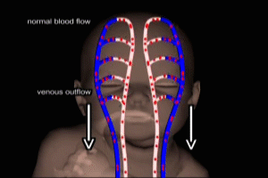
Interruption of adequate blood flow to the fetal brain (hypoxia) may occur during prolonged shoulder dystocia. This is a rare but potentially devastating occurrence, and may result in permanent impairment of higher brain functions, or in extreme cases, fetal death. The exact mechanisms underlying fetal hypoxia during prolonged shoulder dystocia are not well understood, but the the 2 most popular contenders currently are compression of the umbilical cord (interrupting maternal-fetal oxygen transfer), and obstruction of venous outflow from the fetal brain due to compression of the neck in the birth canal.
MATERNAL INJURIES
Severe bleeding after delivery (post-partum hemorrhage) and extension of episiotomy incisions resulting in tears of the rectum (fourth-degree laceration) are the most common maternal injuries related to fetal shoulder dystocia. Other reported complications include cervical and vaginal lacerations, loss of bladder function, uterine rupture, separation of the maternal pubic symphysis, and lateral femoral cutaneous nerve injury related to overzealous use of the McRoberts maneuver (described below).
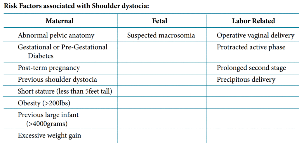
FACTORS INCREASING THE RISK OF FETAL SHOULDER DYSTOCIA
Studies have established a significant relationship between increasing birth weight and the risk of shoulder dystocia. In births not complicated by maternal diabetes, only 5% of infants weighing between 4000 and 4250 grams at birth typically experience shoulder dystocia, while for infants weighing 4750-5200 grams, almost 21% are so affected. Despite these findings, attempts to predict shoulder dystocia based on previous history of over weight births (macrosomia), pre-existing or pregnancy induced non-insulin dependent diabetes, excessive maternal weight gain, and delivery after expected dates, have proven to be unreliable at best. Likewise, fetal birth weight estimates based on ultrasounds performed in the latter stages of pregnancy have not correlated well with the incidence of shoulder dystocia. The most recent recommendation of the American College of Obstetricians is that there is no contra-indication to attempted vaginal delivery for any infant estimated to be up to 5000g (where maternal insulin-dependent diabetes is not present), or up to 4500g for mother with diabetes.
Mothers with insulin-dependent diabetes whose babies are of high birth weight show a significantly higher risk for encountering fetal shoulder dystocia during labor. It is thought that this relates to changes in fetal body proportions, including larger chest and trunk circumferences, larger bisacromial diameters and chest to head ratios, all of which may contribute to inadequate shoulder rotation at the pelvic inlet.
Mothers who have had a prior history of fetal shoulder dystocia during labor should also be considered in a special risk category for recurrence with subsequent pregnancies.
MEASURES TO RELIEVE FETAL SHOULDER DYSTOCIA
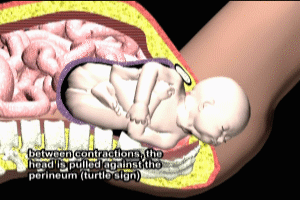
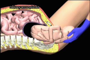
Shoulder dystocia is most often recognized after delivery of the fetal head, when gentle downward traction on the head fails to accomplish delivery. The so-called “turtle sign” is often noted – after the delivery of the head, the head is pulled back against the external vagina (perineum).
Once dystocia is recognized, the mother should be stopped from pushing until the shoulders have been freed. If the umbilical cord is wrapped around the neck (nuchal cord), an attempt should be made to pull it over the head. If this fails, the cord should not be cut, but rather left intact until complete delivery has been accomplished. Fetal shoulder dystocia is considered an obstetrical emergency, and steps should be rapidly instituted to relieve the problem. A delay of more than several minutes between head and shoulder delivery increases the chance of brain damage due to hypoxia.
MCROBERTS MANEUVER
The McRoberts maneuver is generally the first step taken once shoulder dystocia is recognized. The maneuver consists of flexion of both maternal thighs into the abdomen. This action causes a flattening of the lower spine and a subsequent increase in the front-to-back dimension of the pelvis. This change is often enough to permit the shoulders to slip through the pelvic inlet. Care must be taken not to be too aggressive in the maneuver as this may result in maternal injury as described previously.
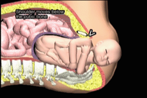
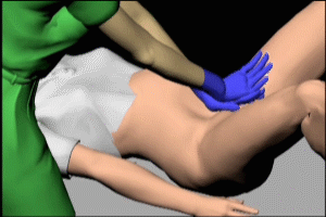
SUPRAPUBIC PRESSURE
Suprapubic pressure is often performed in conjunction with the McRoberts maneuver, or shortly thereafter if the McRoberts is unsuccessful. It consists of downward pressure applied with the heel of the hand directly above maternal pubic bone. The intended effect is to push the impacted shoulder below the pubic bone, thus freeing it to move forward through the inlet. Proper positioning of the hands at a point directly above the pubic bone is critical, as pressure applied at a higher level will compress the uterus and may aggravate brachial plexus stretch or umbilical cord compression and fetal hypoxia.
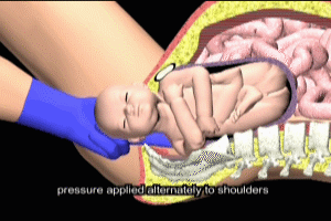
WOODS SCREW MANEUVER
If McRoberts and suprapubic pressure fail to relieve shoulder dystocia, a variety of additional techniques may be attempted, including the Woods Screw and Rubin maneuvers. The Woods Screw consists of application of the operator hands to the anterior and posterior shoulders. Pressure is then applied to rotate the shoulders, moving them into an oblique position, thus relieving the impaction. The Rubin maneuver (reverse Woods) involves application of pressure to the back (posterior) of whichever shoulder is most accessible, again with the goal of rotation into the oblique position.
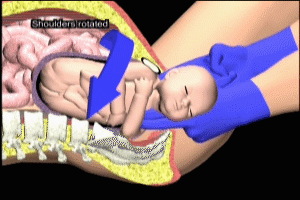
There are many variations in the Woods Screw-type maneuver, with many operators developing their own favorite versions.
POSTERIOR ARM DELIVERY
Delivery of the posterior arm has the effect of pulling the posterior shoulder into the pelvic inlet. When successful, this has the effect of dropping the anterior shoulder beneath the pubic bone. The operator reaches in along the maternal spine, flexes the posterior arm at the elbow, and then delivers the arm with a sweeping motion along the fetal chest.
ZAVANELLI, SYMPHISIOTOMY, HYSTEROTOMY
When the above measures have failed to relieve shoulder dystocia, more aggressive, so-called “heroic” measures may be required. The Zavanelli maneuver consists of an attempt to replace the fetal head back into the labor canal, thus disimpacting the shoulders, and relieving stress on the brachial plexus and umbilical cord. Once accomplished, the decision can be made whether to re-attempt vaginal delivery or proceed to an emergency caesarian section.
Symphisiotomy involves the obstetrician performing an external incision of the symphysis pubis, the ligament that joins the right and left pubic bones. This results in a separation of the pubic bones with subsequent release of the impacted shoulder.
Hysterotomy is the term used for the uterine incison performed during a Caesarian Section, and involves performing a transverse incision along the lower end of the uterus. The posterior arm is delivered through the incision, and the operator then rotates the shoulders to the oblique position to effect delivery.
Cal Shipley, M.D. copyright 2020
