Man Runs a Marathon with a Pulmonary Embolism
(Pulmonary Embolism Review)
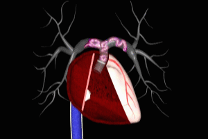
The title of this post sounds kind of cheesy, doesn’t it? It reminds me of when I was a kid, and we use to marvel at the headlines in the “National Enquirer” down at the local candy store, with stories like “Man lives for 2 years without head…” But that’s the amazing thing about the practice of medicine – you run into patients time and again that lend credence to that old saw about truth being stranger than fiction. And the title of this post is true, or at least, it could be. I guess I had better explain…
WHAT IS A PULMONARY EMBOLISM?
For the uninitiated, simply put, a pulmonary embolism is an abnormal clot of blood present in the arterial side of the blood circulation in the lung(s). I use the term “abnormal” because not all blood clots in the pulmonary circulation are problematic. In fact, small clots are constantly being formed throughout our bloodstreams 24/7. As soon as these clots are formed, naturally generated “clot-busting” chemicals rush to the affected spot and dissolve the clots, so that any interruption in blood flow through the affected artery is kept to a minimum. This clotting and de-clotting process is just another of the numerous examples of a balanced system within our physiology. In the case of a pulmonary embolism, however, the clot, or clots are large enough, or numerous enough, that the natural clot-busters can’t keep up with the demand to dissolve. This results in a prolonged interruption of blood flow through the affected artery or arteries. In the lungs, this interferes with the transfer of oxygen from the airways (bronchial system) to the bloodstream.
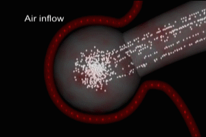
The greater the total amount of abnormal clot present (“clot burden”), the greater the interference with the oxygenation process. Healthy children and adults have a fairly large respiratory capacity – at any given moment, we are using only a fraction of our bodies capacity to absorb oxygen. As a result, most of us can tolerate the presence of smaller amounts of abnormal clot in the pulmonary arterial system with minimal or sometimes even no obvious symptoms. As the amount of clot increases, signs of lowered blood oxygen levels (hypoxia) appear – primarily shortness of breath very often associated with chest pain. The onset and severity of the symptoms is also a function of the level of fitness and physical activity of the affected individual – given the same clot burden, and all other things being equal, a couch potato is less likely to notice symptoms than a marathon runner.
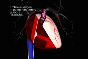
THE MARATHON RUNNER
Which brings me to the subject of today’s post – the marathon runner. In addition to running my medical legal graphics business Monday to Friday, I still see patients at a local Urgent Care center on weekends. Some months ago, a 25 year-old man presented to our clinic with a complaint of shortness of breath and chest pain. Let’s call him “Fred”. Fred had noted the shortness of breath for about a week prior, but had just developed the chest pain 24 hours before coming to see me. He was a long-time runner, logging about 40 miles per week, and had just completed the Portland marathon 3 weeks prior to his clinic visit. He had been scheduled to run a half-marathon right around the time his shortness of breath developed, and found that he could complete less than a mile, and then had to stop because he felt so short of breath. He had been under a lot of pressure at work recently, and thought perhaps his shortness of breath was stress-related. But when he developed fairly sharp, persistent pain in the center of his chest 24 hours prior to his visit, he decided to get it checked out.
PULMONARY EMBOLISM: THE HISTORY
Fred had no history of previous serious medical problems. Specifically, he had no history of clotting disorders, nor of abnormal clots anywhere in his body, was on no medications, and had never been a smoker. Nor did his family members have any such history. He denied any pain or swelling in his legs (a common source of pulmonary embolism is clots that form in the deep veins of the legs – these are called deep venous thromboses or DVT (see Well’s DVT probability table here and causes and diagnosis of DVT here). The only point of interest in his history was that he had come to his regular doctor about a month prior (1 week before running the marathon) with a complaint of mild central chest discomfort on exertion that had been present for several days. This was not interfering with his marathon training, but with just 1 week to go before his marathon run, he was being extra cautious about any bodily changes, and wanted to nip any possible problem in the bud, lest it stop his run (I’ve been there – I ran my one and only marathon in the 80′s, and as the run approached, I was sure every little ache and pain was about to become something big!) On examination that day, his doctor was able to demonstrate some tenderness in Fred’s anterior chest wall, but the exam was otherwise normal. A chest x-ray and EKG looked perfectly normal as well. His doctor concluded it was some mild irritation in the chest wall, and prescribed some anti-inflammatory medication, which Fred stated seemed to do the trick. By the day of the marathon, the chest discomfort was resolved.
THE PHYSICAL EXAMINATION
On examination, he was a lean, fit appearing young man – he looked like someone capable of running 26.2 miles. He was clearly anxious about his current symptoms, and was close to tears when discussing the stress he had been under at work. I think he felt like a fraud, and that his symptoms were being generated by his anxiety. Fred’s physical examination was completely normal. his vital signs – blood pressure, pulse, and temperature were all normal, as was the oxygen saturation (pulse ox) in his blood – 99% on room air – anything at 96% or higher is considered normal). His lungs were clear when I listened with my stethoscope, as were his heart sounds and pulse rhythm. His lower limbs showed no sign of unusual swelling or tenderness – the kinds of findings we would see with deep venous thrombosis (DVT). In fact, the most interesting aspect of his physical examination was what wasn’t present – try though I might, I could not elicit tenderness when I pressed on Fred’s chest wall. This was the opposite of his examination a month prior, when his own Doc had demonstrated tenderness, and it left me with a fairly large problem: what was causing Fred’s chest pain?. Click here to view Well’s Criteria for assisting in the diagnosis of pulmonary embolism.
PULMOMARY EMBOLISM: THE DIFFERENTIAL DIAGNOSIS
So here was a seemingly very healthy 25 year-old man, who nevertheless had a 1 week history of shortness of breath – severe enough to prevent him from running, and a 24 hour history of sharp chest pain, for which my physical exam had yielded no clues as to its origin. Based on all of this, I had fairly rapidly formulated a list of possible diagnoses for Fred – a so-called “differential diagnosis”. That’s a term doctors use when they’re not absolutely certain what the diagnosis is – this happens a good percentage of the time – (Medicine is, as yet, still an inexact science). The “differential” gives us a list of possible diagnoses. We then take further steps to attempt to eliminate the diagnoses on our list, until we are (hopefully) left with the one, true, diagnosis. Of course, it doesn’t always work that simply, but you get the idea. Once we have created a differential diagnosis, the steps we take generally involve some type of testing, be it blood tests, imaging studies, etc. Sometimes, we discuss the case with someone smarter than us (we call these smarter people “specialists”) to help guide us along the way.
But I digress! My differential diagnosis for Fred that day came down to three possibilities:
1) myocardial infarction (heart attack) – not impossible, but extremely unlikely in a fit 25 year old – I’ve never seen an MI in one so young in 30 years of practice
2) pulmonary embolism – again, very unlikely in a fit specimen like Fred, with no risk factors, no personal history of similar problems, and no family tendency to such problems.
3) anxiety – really? Can anxiety cause sharp chest pain? Absolutely! I’ve seen this dozens of times. What about shortness of breath? Well, yes, I’ve definitely seen shortness of breath in anxious folks, but typically, this occurs during a special kind of very acute anxiety called a “panic attack”. Prolonged shortness of breath, persistent over several days is a bit unusual for anxiety.
THE DIAGNOSIS
So – 3 possible diagnoses to sort through – what steps to take to nail down the one, true diagnosis? When anxiety is on the differential list, the only way to make the diagnosis is to rule out all of the other diagnoses on the list first. If they all turn up negative, then you’re left with anxiety. This is known as a “diagnosis of exclusion”. Exclude all the other diagnoses, and you’re left with the diagnosis that hasn’t been excluded. Sounds a bit unscientific, but, unfortunately, there’s no blood or imaging test one can perform that will positively show that a physical symptom is related to anxiety.
Ruling out a myocardial infarction (heart attack) is relatively easy – get an EKG (electrocardiogram – I’ve never been sure where the “K” came from!) and test Fred’s blood for cardiac enzymes – these are enzymes that are released when fibers of the heart muscle (myocardium) are damaged or die, as in a heart attack.
PULMONARY EMBOLISM AND THE D-DIMER TEST: THE DOUBLE-EDGED SWORD
Ruling out a pulmonary embolism is a bit trickier proposition. These days, if we suspect pulmonary embolism, we do a blood test for “D-Dimer”. D-Dimer is a protein fragment that occurs as a product of the activity of natural clot-busters on clots. The larger the clot, the greater the level of D-dimer detectable in the bloodstream. The difficulty with D-dimer is that it lacks a high degree of specificity – that is, there are processes in the human body that can give rise to an elevated level other than the abnormal clots we see in pulmonary embolism. This may give rise to falsely positive (ie elevated) D-dimer levels. On the other hand, the sensitivity of the test is very high. High sensitivity means that there is a very low rate of falsely negative results with D-dimer. That is, if the levels are not elevated, you can bet your bottom dollar that there is not an abnormal clot process occurring anywhere in the body (including the lungs), because the D-dimer test is very sensitive to the presence of abnormal amounts of clot.
So if the D-dimer is normal, hallelujah, we’ve ruled out a pulmonary embolism. The problem arises if the test is elevated, even slightly elevated. Not only have we not ruled out a pulmonary embolism – now we have to take a further step to test for it. Currently, that further step is a CT (Computed Tomography) scan of the lungs. The CT can show us definitive proof of the presence or absence of pulmonary embolism. Why not just do the CT in the first place, you ask? For one thing, to get the best imaging results, CTs are generally done after an injection of contrast into the patient intravenously. A small percentage of patients may have a significant allergy reaction or intolerance ot the contrast. Secondly, and of greatest significance for all who undergo a CT, is the high amount of radiation exposure the CT involves. A CT of the chest is delivers the radiation equivalent of 200 plain film chest x-rays! That’s a significant amount of radiation, particularly in individuals who, for one reason or another, have to have repeated CTs over the course of their lives. And where there is more radiation, there is more cancer. It is now estimated that up to 2% of all cancers in the United Sates annually are related to imaging study radiation exposure.
So when a physician orders a D-dimer, knowing that the test carries with it a significant probability of a falsely positive result, she is acknowledging, up front, that the patient involved may end up having a CT scan, and therefore, be exposed to high levels of radiation. For this reason, we try to limit D-dimer testing to patients in whom there are symptoms, and/or risk factors, that increase the pre-testing chance that they may have a pulmonary embolism. And because each radiation exposure adds to the previous exposures over a lifetime, we are particularly reluctant to use the CT scan in younger patients without a really good reason for doing it. Here’s a link to a recent study published in the Journal of the American Medical Association involving D-dimer testing to rule out pulmonary embolism.
FRED SAYS “ENOUGH TALK! PLEASE DIAGNOSE ME ALREADY!”
Ok Fred – what am I going to do with you? You’re 25 years old, you’ve had shortness of breath for a week, and chest pain for a day, and you have no risk factors for pulmonary embolism. Not only are you low risk for pulmonary, embolism you’re an athlete – you’re not sitting around all day waiting for the blood in your veins to clot, you’re out pushing that blood around your system, decreasing the chance of abnormal clotting. You have a completely normal physical exam, and you’re oxygenating at 99% of your bloodstream’s capacity (not a finding you would typically correlate with pulmonary embolism). And you’ve been under a lot of stress lately, and we know that stress can cause just about any physical symptom in the book. All of these considerations are racing through my mind after I’ve spoken to and then examined, Fred. How would I decide whether or not to risk exposing this very young man to a potentially needless dose of CT radiation?
Well, in the end, I did order the D-dimer test, and I did it for 3 reasons: First, even taking into account his recent anxiety, Fred’s shortness of breath seemed inappropriately severe and persistent to be blamed on on a case of nerves. Second, I had no good explanation for his chest pain. The previous diagnosis of chest wall pain a month prior just didn’t apply based on his current exam. And even before I saw his EKG, I knew that a myocardial infarction (heart attack) was extremely rare in a fit young individual. And third, something in my gut told me that there was more to Fred’s current physical problems than anxiety. Call it intuition if you will, but after 30 years of practice, there are just times when you feel that something significant is going on in a patient, even though you can’t put your finger on exactly what it is, or why you feel that way. And that’s how I felt about Fred that day in clinic.
THE PAYOFF
Fred’s chest xray and EKG were normal, as were all of his blood tests, with the exception of the D-dimer. The D-dimer level was 3300 (normal at our lab is 500 or less). So off to CT we bundled him for a lung scan, and lo and behold, his pulmonary artery system was full of clots on both the left and right side. He was promptly transferred to the Emergency Department, and subsequently admitted to hospital for anit-coagulation therapy. My gut feeling paid off – this time…
TIMELINE
In considering Fred’s very unusual case after the fact, I found myself dwelling on one question: how long had Fred been walking (or running) around with his pulmonary embolism (or emboli as it turned out)? I’m 100% certain that he’d had the lung clots for at least the week prior to his visit with me – the period of time when he had noted the marked shortness of breath on exertion. But wait, he’d visited his own physician a month before with complaints of chest pain. Might he have had some degree of pulmonary embolism then? And if so, how is it possible that he could have completed a marathon run just 1 week later. One would presume that, with any degree of blood flow compromise in his lungs due to embolism, he’d have perceived some kind of problem with his endurance.
RECANALIZATION IN PULMOMARY EMBOLISM
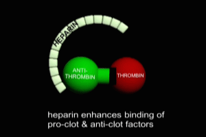
Actually, it is possible that Fred could have had chest pain due to a pulmonary embolism, and then run a marathon 1 week later. Earlier in the post, I touched on the ongoing balance between small clots forming in the body, and naturally occurring clot-busting chemicals that dissolve the clots to keep us out of harms way. When an abnormal clot, such as a pulmonary embolism, initially occurs, the clot-busters in our body go to work, just as they would on a smaller clot, and in the early stages of the embolism, the clot-busters may actually succeed in re-opening blocked arteries. This process is known as recanalization, and it can permit an individual with pulmonary embolism to have initial symptoms, followed by periods of apparently normality. Unfortunately, particularly where there is a process feeding the pulmonary embolism, such as clots in the legs (DVT) that repeatedly break off and travel via the venous system to lodge in the pulmonary arteries, eventually, the total amount of abnormal clot (clot burden) simply becomes more than the clot-busters can cope with, and the affected individual inevitably experiences significant symptoms, or even death.
This process of recanalization may well have been the factor that allowed Fred to run a marathon after an initial pulmonary embolism. I’ll never know for sure whether he had the embolism that week prior to the marathon. It could have simply been a coincidence – some minor chest wall inflammation a week prior to the marathon, then onset of pulmonary embolism sometime after. But coincidences in medicine have always made me uncomfortable. My experience has been that events in a patient’s medical condition that appear coincidental, more often than not, turn out to be related.

DENOUEMENT
Fred’s doing well now. Six weeks after diagnosis, he is still on anti-clot medication, but is back training for another marathon. Even after a very extensive workup, we never did figure out what caused his pulmonary embolism. This inability to pin down the underlying cause of embolism is unusual but not rare. My guess is that he’ll be returning to the clinic very promptly should he ever develop any kind of chest pain or shortness of breath in the future. For an recent comprehensive clinical review of pulmonary embolism in the New England Journal of Medicine, click here.
RELATED VIDEOS
Cal Shipley, M.D.
